Bulging Disc vs Herniated Disc: How Each Shows on MRI, What Symptoms Mean, and When to Get Evaluated.
Disc bulges appeared in 52% of adults without back pain in an MRI study described by the North American Spine Society. That's a useful starting point if you've opened an imaging report and wondered whether a bulge explains everything you're feeling.
The bulging disc vs herniated disc distinction describes anatomy, not a pain score. To understand what your report means for you, a clinician needs to connect the picture with where your symptoms travel, how you're moving, and whether a nerve is losing function.
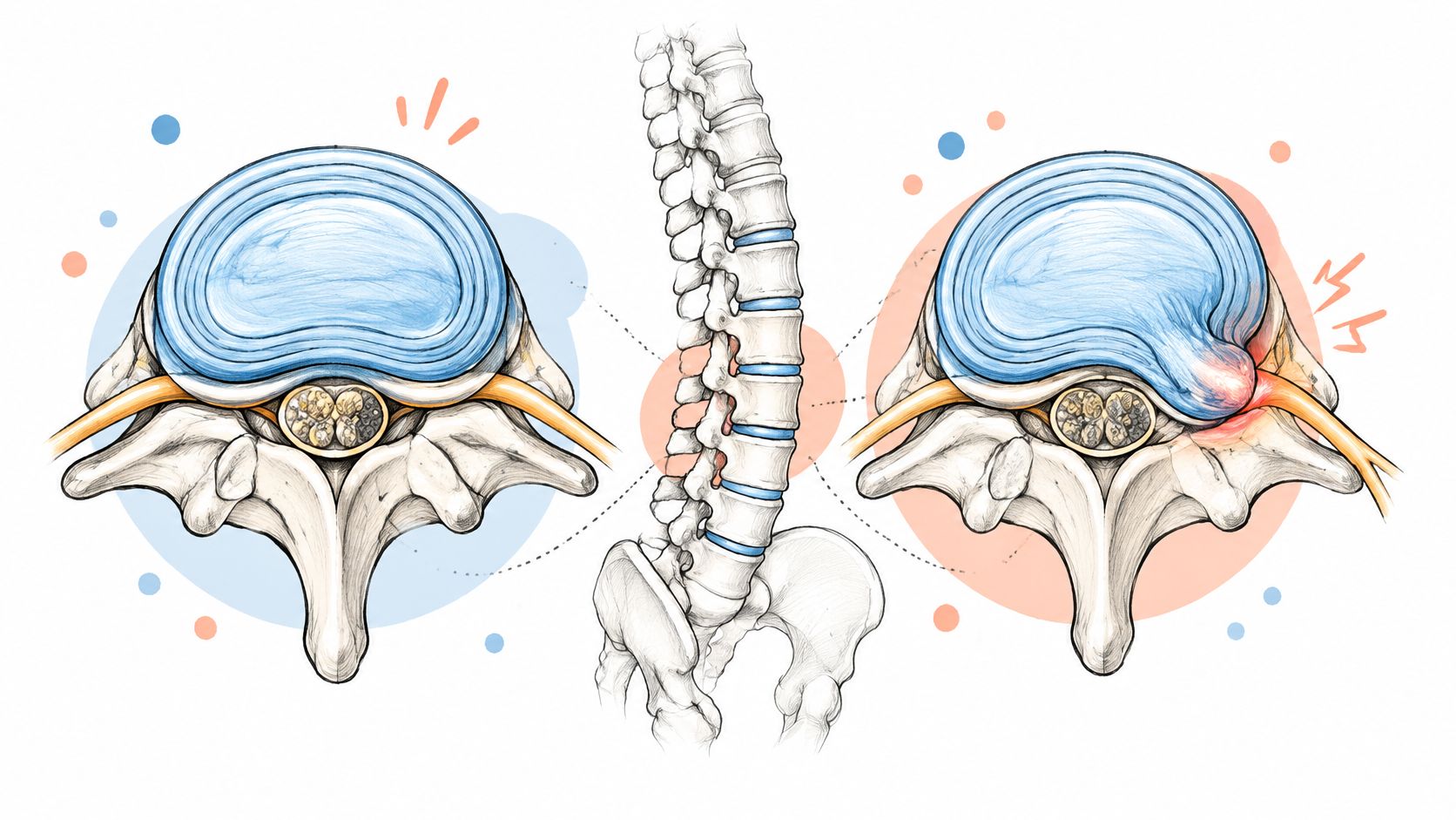
A disc bulge extends beyond the disc's usual boundary over a broad area. A herniation describes a more localized displacement of disc material through a defect in the outer ring, called the annulus. These terms describe the shape of a finding on imaging; interpreting its significance requires looking at the whole report and the clinical picture.
The American Association of Neurological Surgeons' herniated-disc guide explains how displaced material can irritate nearby nerves. The shape of a finding alone does not establish what is causing symptoms.
| Criterion | Disc Bulge | Disc Herniation |
|---|---|---|
| Definition | Broad extension of disc tissue beyond the normal margin | Focal displacement of disc material through an annular defect |
| Share of disc circumference | Broad area of the disc edge | Localized area of the disc edge |
| Nerve contact | May occur, depending on location | May occur, depending on location |
| Typical symptoms | Often none; back or neck pain can occur | Symptoms vary; pain, numbness, tingling, or weakness can occur if a nerve is involved |
| Usual cause | Often associated with age-related changes | May occur with disc degeneration and an annular defect, sometimes after a strain |
| Subtypes | Not commonly divided into subtypes | Protrusion, extrusion, sequestration |
Circumference describes how widely the finding extends around the disc. It says little about how much pain someone will feel. A broad change may be away from a nerve, while a localized displacement may be near one.
A herniation can also irritate a nerve through inflammation around the nerve root. The resulting symptoms may involve both local contact and inflammation.
Protrusion, extrusion, and sequestration are types of herniation. In a protrusion, the base of the material extending outward is relatively broad. An extrusion extends beyond a narrower connection, and sequestration means a fragment has separated from the disc.
These terms describe the material's shape and connection to the disc. They help characterize the MRI finding, while treatment decisions depend on the person's symptoms and examination.
Find the exact wording in your MRI report: bulge, protrusion, extrusion, or sequestration. Note any mention of nerve contact, displacement, or compression as well. Together, those details give a fuller description of the scan.
Back pain and nerve-root pain can overlap, but they aren't interchangeable. The most useful clues concern where symptoms travel and whether sensation or strength changes, rather than whether the pain feels severe.
Localized back or neck pain that changes with posture, stiffens after sitting, and eases with comfortable movement can fit a mechanical pain pattern. Muscles, joints, and discs may all contribute, so this pattern doesn't identify a particular structure.
Pain traveling down an arm or leg in a relatively consistent band raises a different question: is a nerve root irritated? In the lower back, this may feel like pain through the buttock and into the leg. In the neck, symptoms may extend toward the shoulder, arm, or hand.
Our guide to nerve pain management offers additional context for recognizing and discussing these symptoms.
Trouble lifting the front of your foot, repeatedly catching your toes, or losing grip strength deserves more attention than pain intensity alone. A clinician needs to distinguish true muscle weakness from movement limited because it hurts.
Say your back feels less painful, but your foot starts slapping the ground when you walk. That isn't reassuring improvement overall. New or worsening loss of function requires medical assessment, even when pain is settling.
Track what you can do, not just how much it hurts. Walking, gripping, lifting the foot, and sleeping comfortably give your clinician different kinds of information.
For nonurgent symptoms, keep a two-week symptom log to bring to your appointment. Record location, triggers, episode duration, numbness, perceived weakness, and whether symptoms are improving or worsening. Don't postpone an evaluation to finish the log if weakness develops or symptoms escalate.
Age-related scan findings are common in people who have no symptoms. A systematic review of imaging in asymptomatic individuals reported the age-specific estimates below, including bulges in approximately 30% at age 20 and 84% at age 80.
| Age | Disc bulge | Disc protrusion | Disc degeneration |
|---|---|---|---|
| Age 20 | About 30% | 29% | 37% |
| Age 80 | 84% | 43% | 96% |
| Older than 70 | More than 75% | No separate estimate stated for this age group | No separate estimate stated for this age group |
The older-than-70 bulge estimate comes from another synthesis described in the clinical discussion of asymptomatic imaging findings, which also reports bulges in about 20% of young adults. These estimates use different age groupings and shouldn't be treated as one continuous dataset.
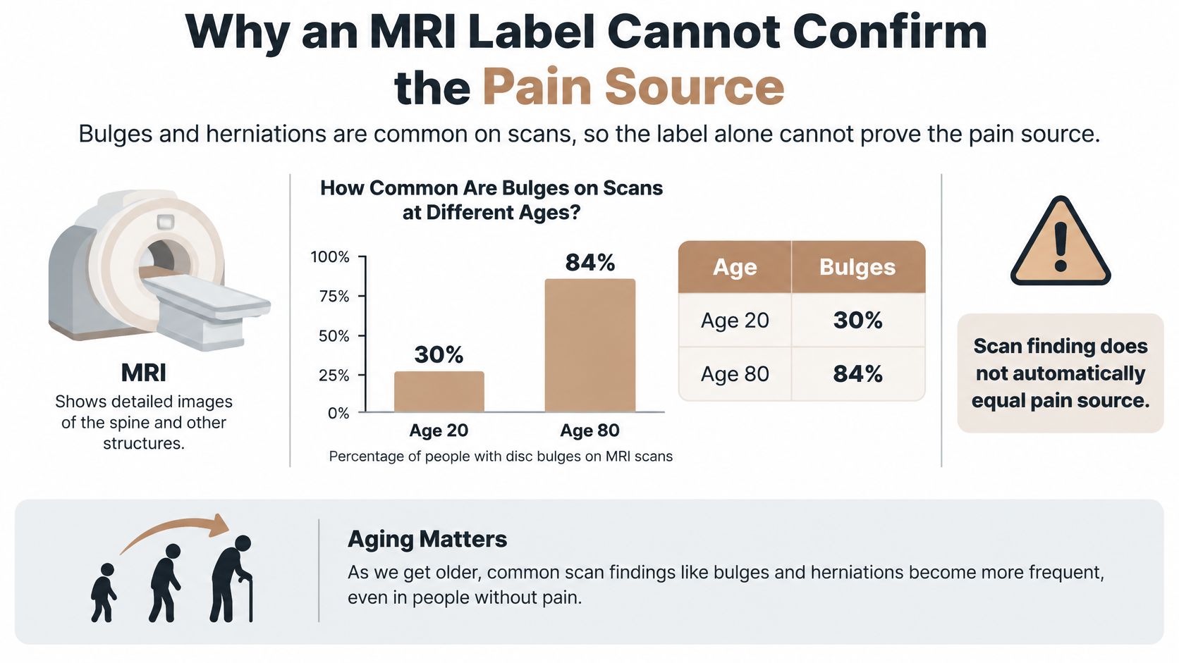
An abnormal scan may therefore describe age-related anatomy rather than explain current pain. That doesn't make your pain imaginary. It means the clinician still has to identify which findings matter.
A bulge without matching nerve findings generally supports conservative, function-focused care and reassessment. A focal herniation accompanied by a matching pattern of limb pain, sensory loss, altered reflexes, or weakness calls for closer neurological monitoring and possibly specialist referral.
For example, a left-sided finding is less convincing as the explanation for exclusively right-sided symptoms. The clinician should explain that mismatch instead of assuming every abnormality needs treatment.
Practical rule: A scan finding becomes more meaningful when its location, side, and affected nerve agree with the symptoms and examination.
Reports may also use terms such as degeneration or spondylosis. Dream Body Clinic's resource on understanding spondylosis on an MRI report can help you distinguish that broader terminology from a specific disc displacement.
Deciding when to get an MRI for back pain is a separate clinical decision from interpreting one you've already had. A bulge isn't automatically less serious than a herniation, and treatment shouldn't follow the most alarming word on the report.
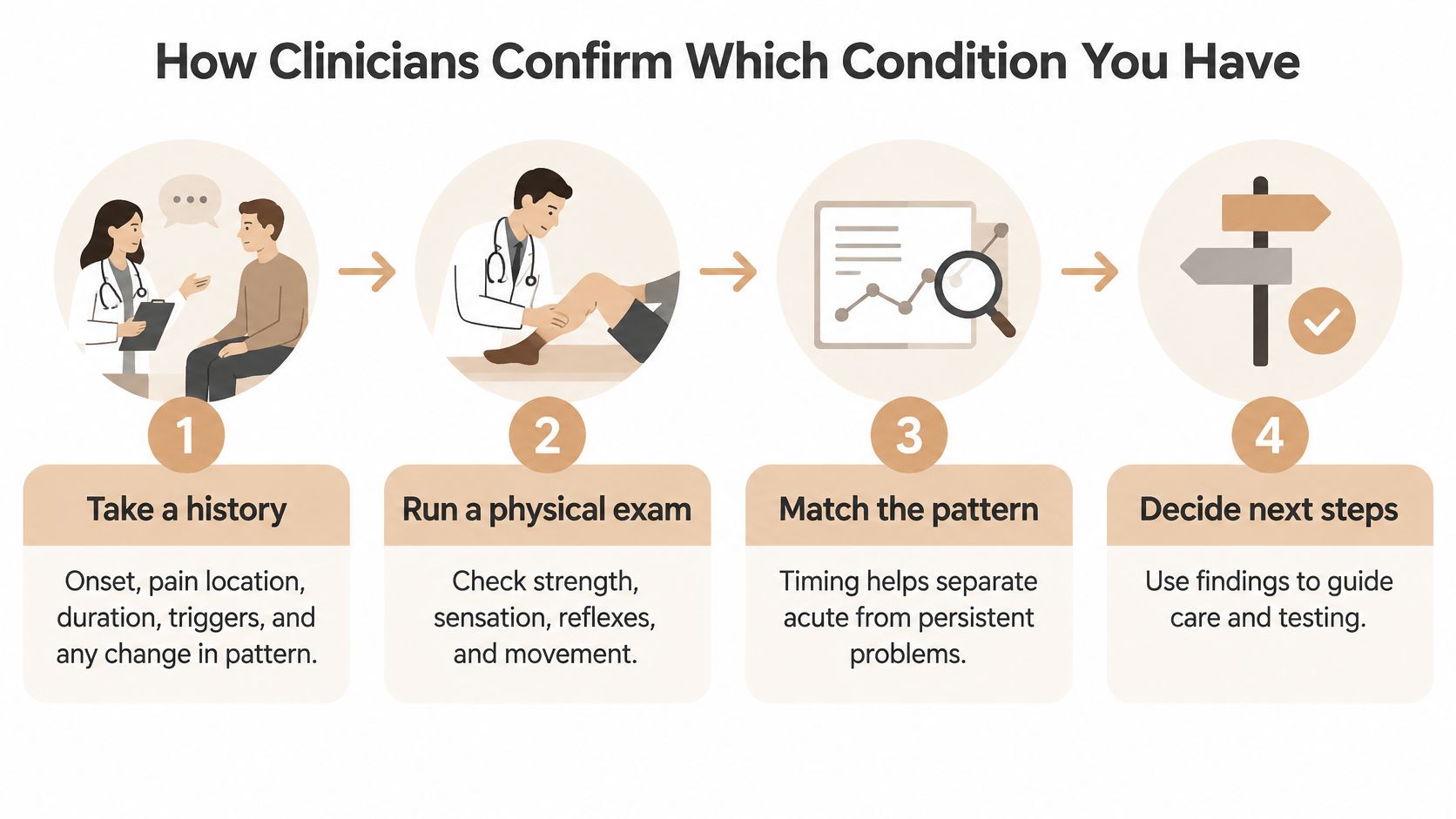
A disc label is one piece of a diagnosis, not the diagnosis itself. Clinicians combine your symptom history, physical examination, and any imaging that could help answer a specific question. The aim is to understand how the condition affects you and decide what care is appropriate.
Take a history to understand how symptoms behave. Describe when they began, where they travel, how long they last, and what makes them better or worse. Mention any change from your usual pattern, especially after an injury.
Check nerve function, since pain alone cannot show how a nerve is working. The clinician assesses muscle strength, sensation, and reflexes, comparing findings between sides. For lower-back symptoms, a straight-leg raise may help determine whether a position reproduces familiar radiating pain rather than stretching a tight hamstring.
Choose imaging to answer a clinical question. At Aspen Falls Wellness, an X-ray or MRI may be suggested during the initial examination to help confirm your condition. X-rays show bones and alignment; MRI provides detail about discs and nearby nerves. Imaging is not automatic at every first visit.
Resolve conflicting findings before settling on a diagnosis. If the examination and imaging point in different directions, the clinician may reconsider other explanations, gather more information, or monitor how symptoms change. A repeat scan may be considered if new or worsening symptoms raise a question that could affect care.
Set a reassessment plan. Identify which symptoms, movements, or strength findings will be checked again. Progressive weakness or bowel or bladder changes call for urgent medical assessment rather than waiting for a routine follow-up.
Refer onward when the problem needs care beyond the clinic's scope. Significant neurological changes, suspected emergencies, or persistent disabling symptoms may require assessment by a medical or surgical specialist.
At Aspen Falls Wellness, Dr. Jeremy Kaier specializes in treating herniated discs, stenosis, and sciatica. An initial assessment helps determine whether an individualized non-surgical plan fits your presentation or whether further evaluation is needed.
Bring the imaging report and, if available, the images themselves. Share what treatments you have already tried and how you responded. This helps the clinician distinguish options that have not been attempted from those that aggravated symptoms or did not help.
A disc herniation can shrink without surgery, and some bulges also regress. A label such as “bulge” or “herniation” does not predict how quickly you will recover. Symptoms, nerve function, and the type of disc change all matter.
Resorption is the body's process of clearing displaced disc material. A 2024 systematic review covering 2,199 cases reported spontaneous resorption in 76.6% of cases, with individual studies ranging from 20% to 96.2%. Resorption occurred mainly during the first 3 to 6 months of conservative treatment.
Ruptured or extruded herniations regressed more often than contained herniations. Material that has moved beyond the disc's usual boundary may be more accessible to the body's immune and vascular responses.
A systematic review of disc imaging findings reported regression in approximately 13% of bulging discs, 41% of protrusions, 70% of extrusions, and 96% of sequestrations. These figures describe imaging changes across groups, not the outcome an individual can expect.
A larger-looking herniation may sometimes shrink more readily than a smaller, contained one. Size alone does not show whether nerve function is at risk.
Pain can ease before a follow-up scan shows a clear change. The reverse can happen too: a herniation may shrink while strength, sensation, or everyday function still need time to recover.
The Merck Manual's reference on lumbar disc herniation describes symptoms easing as a herniation dries out and may reabsorb over roughly 6 to 9 months. That biological process can continue after initial pain relief begins. For more detail on these different milestones, see how long herniated disc recovery can take.
A wait-and-see approach means tracking how symptoms and function change. Improving walking tolerance, stable strength, and fewer disruptions to daily activities can support continuing a plan. Worsening function calls for reassessment, whatever the scan shows.
For uncomplicated acute radicular symptoms, care often begins with adjustments to daily activity, appropriate medication, and physical therapy. A 2024 clinical review of lumbar disc herniation reports that many symptomatic episodes resolve within approximately 6 to 8 weeks. Serious neurological deficits can change the treatment approach.
| Option | Typically Used When | Typical Timeline |
|---|---|---|
| Activity modification and medication | First-line for most acute radicular pain | Many symptomatic episodes resolve within about 6 to 8 weeks |
| Physical therapy and stabilization exercise | Symptoms persist after initial activity modification | Timing is individualized and reviewed as symptoms and function change |
| Non-surgical spinal decompression | Selected disc-related pain needing additional conservative support | Course length set by the clinician |
| Laser therapy and massage | Adjunct care targeting inflammation and soft-tissue pain | Response reviewed during the individualized treatment course |
| Epidural steroid injection | Severe radicular pain not improving | Follow-up set by the treating medical clinician |
| Microdiscectomy surgery | Progressive weakness, cauda equina symptoms, or failed conservative care | Faster short-term relief than conservative care; similar long-term results in selected patients |
A systematic review comparing treatments for lumbar disc herniation with radiculopathy found better short-term outcomes with microdiscectomy than with advice or conservative care, while long-term outcomes were equivalent. It also found support for stabilization exercise compared with no treatment.
Activity modification involves reducing loads that aggravate symptoms while keeping up tolerable movement. Medication choices should account for your medical history and other medicines.
Physical therapy and personal exercise plans can help rebuild movement tolerance and strength. Exercises may need adjustment if they increase traveling pain or bring on new neurological symptoms.
Aspen Falls Wellness offers non-surgical spinal decompression using the DRX9000. A customized treatment plan may include chiropractic adjustments, DRX9000 Decompression, and/or MLS Laser Therapy.
Patients typically begin to experience back pain relief within weeks in Aspen Falls Wellness's care, though individual results vary. Treatment tolerance, neurological findings, and functional improvement help guide whether to continue or change course.
Improving neck pain without weakness usually calls for a planned evaluation and routine follow-up. A scan label alone does not set the urgency. The pattern of symptoms, changes in strength or sensation, and day-to-day function help guide what to do next.
A systematic review of conservative management for lumbar disc herniation summarizes guidelines that commonly recommend at least 6 weeks of conservative care before elective surgical decompression is considered, when motor weakness and cauda equina symptoms are absent. This is a treatment checkpoint, not a guarantee of recovery or a reason to wait if symptoms worsen.
| Situation | Likely Next Step | Urgency |
|---|---|---|
| Back or neck pain, no leg symptoms, improving | Conservative care and routine reassessment | Routine follow-up |
| Radiating leg or arm pain, no weakness | Conservative care with monitoring for about 6 weeks, as described in guideline summaries for lumbar disc herniation | Schedule an evaluation |
| Radicular pain with numbness or reflex change | Neurological exam and specialist referral if not improving | Prompt evaluation |
| Symptoms unchanged after 6 weeks of care | Review the plan and discuss whether a surgical consultation is appropriate | Schedule an evaluation |
| Progressive weakness or difficulty walking | Urgent medical assessment | Urgent |
| Bowel or bladder changes, saddle numbness, or suspected cauda equina symptoms | Emergency evaluation | Emergency now |
The six-week timing applies to lumbar herniation care. Arm symptoms call for an assessment of the neck, not automatic use of a lower-back treatment schedule.
Radiating pain without weakness still warrants an examination, especially if it limits sleep, work, or walking. Persistent numbness or an abnormal reflex gives the clinician a neurological finding to document and monitor. If progress stalls, the diagnosis and care plan may need review, including whether specialist input is appropriate.
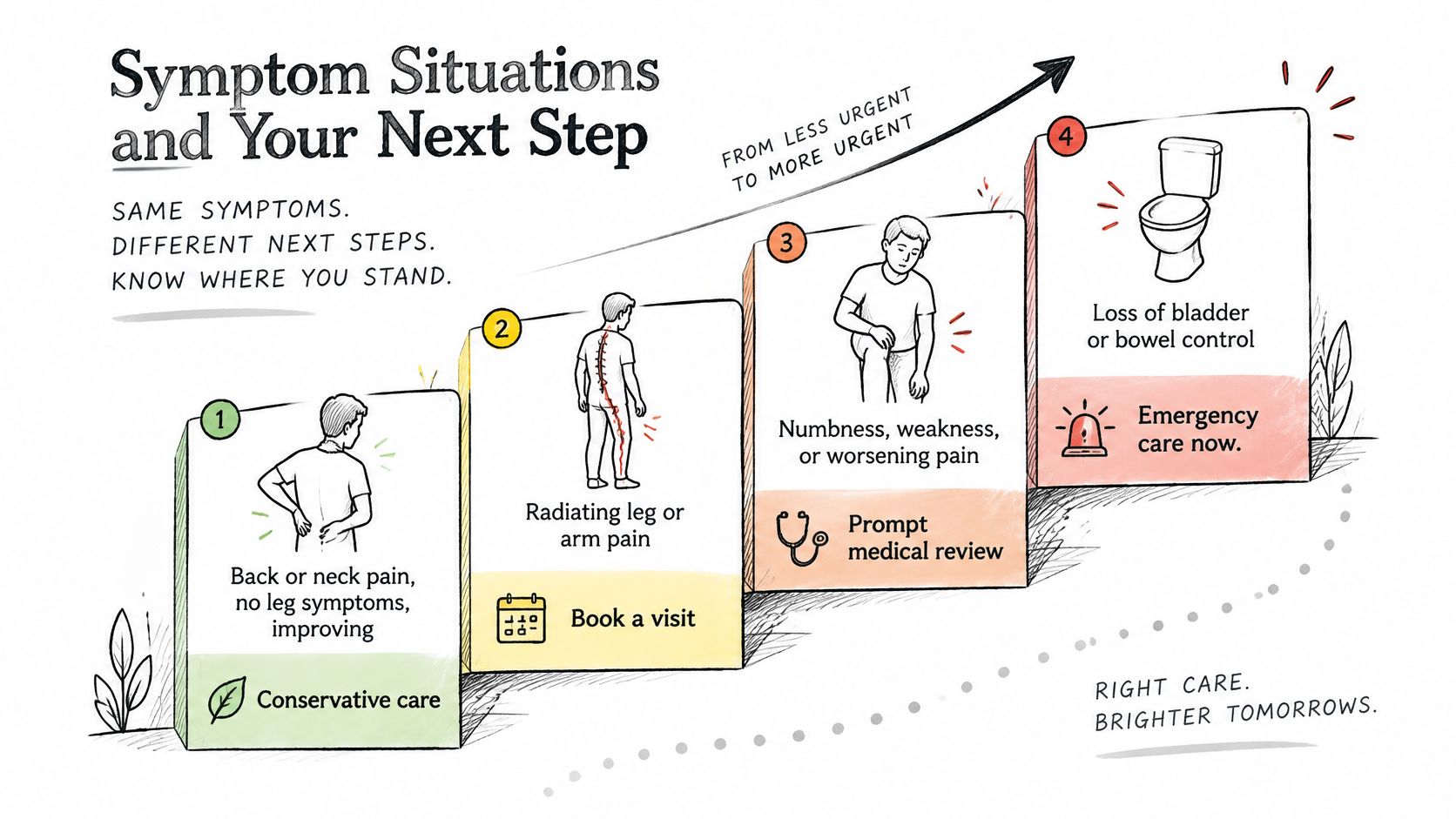
Progressive weakness or new difficulty walking needs urgent medical assessment. New urinary retention, loss of bladder or bowel control, or numbness around the groin and inner thighs can indicate cauda equina syndrome, an emergency involving nerves in the lower spinal canal. Seek emergency evaluation rather than waiting for a routine appointment.
If you have no emergency signs, prepare for an examination instead of choosing treatment from the MRI report alone.
At Aspen Falls Wellness, an individualized plan may include chiropractic adjustments, non-surgical DRX9000 spinal decompression, MLS Laser Therapy, or rehabilitation exercise, depending on the diagnosis. The practice can refer patients to external specialists when their needs fall outside its in-house scope.
A useful assessment connects the scan to your symptoms and examination findings, then sets out what progress to watch for and when the plan should change.
Aspen Falls Wellness offers individualized assessment and non-surgical care for disc-related symptoms, including DRX9000 spinal decompression, chiropractic care, and rehabilitation exercise. Visit Aspen Falls Wellness to arrange an initial examination in Salt Lake City or Sandy and bring your MRI report to discuss how its findings relate to your symptoms and next steps.

Stay updated with the latest chiropractic news and exclusive offers.