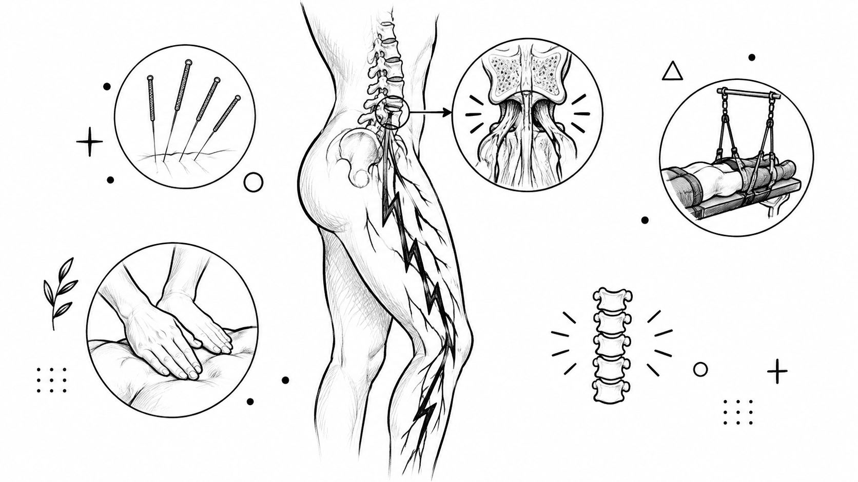
When to get an MRI for back pain: emergency red flags, the 6-week conservative trial, insurance rules and what an MRI can and can't show.
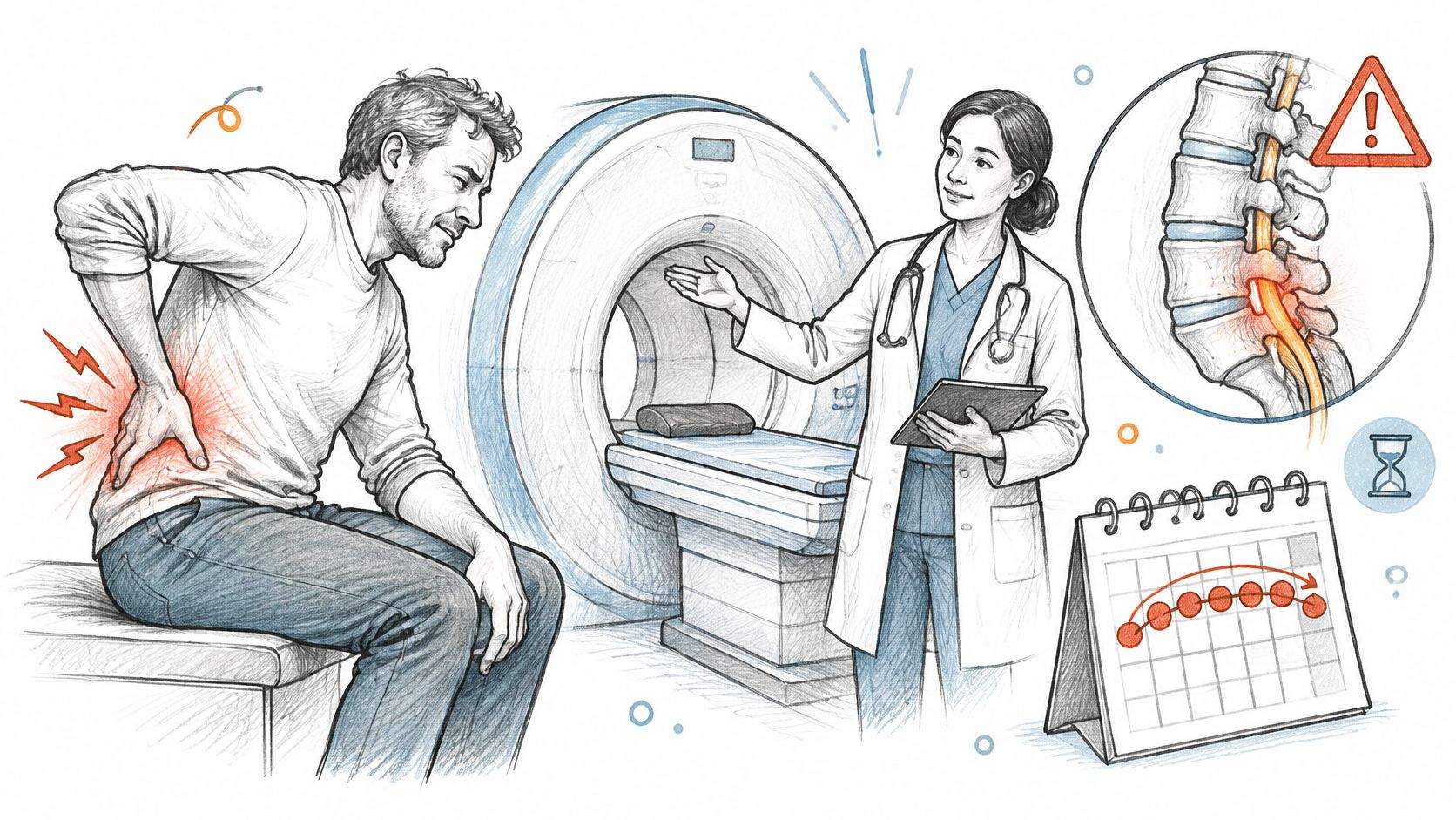
For uncomplicated back pain with no warning signs, an MRI is generally not recommended during the first 6 weeks, but bladder or bowel changes, numbness in the groin or inner thighs, or progressive leg weakness need same-day medical assessment. You're probably reading this because your back still hurts, you can't sit through a drive or sleep through the night, and you want a picture that finally explains why. That urge makes sense. Pain that won't settle creates a simple question, when to get an MRI for back pain, and a frustrating answer from many clinicians: not yet.
The useful way to decide isn't to stare at a calendar. It's to ask one question at each visit: would a scan change the plan right now? If the answer is no, documented care and a recheck are usually safer and more useful. If the answer is yes, because nerve signs are worsening or an injection or surgery is being considered, the scan earns its place.
For most adults with back pain and no warning signs, an MRI isn't the first step, and timing depends on how your symptoms behave over time. Uncomplicated pain under six weeks usually gets structured conservative care with a recheck, not a scan.
Find the situation that matches you most closely. That row sets your clock, not your pain score.
| Situation | MRI timing | Next step |
|---|---|---|
| New bladder or bowel loss, or numbness in the groin or inner thighs | Emergency, same day | Go to an emergency department now |
| Progressive leg weakness or foot drop | Urgent, within days | Urgent spine or neurology evaluation |
| Suspected cancer, infection, fracture or major trauma | Expedited | Same-week medical coordination |
| Uncomplicated pain, no red flags, under 6 weeks | Not routinely recommended | Structured conservative care with a recheck |
| Leg pain (sciatica) persisting past 6 weeks with little improvement | Reasonable to consider | Clinician checks whether the result would change treatment |
| Function loss despite 6 weeks of care, with injection or surgery under consideration | Reasonable to order | Specialist referral with imaging |
| Pre-injection or pre-surgery planning | Indicated | Ordered by the treating specialist |
Pain intensity alone rarely moves a case to MRI. A person rating pain as severe but with normal strength, sensation, and reflexes and steady improvement usually stays in the conservative row. A person with milder pain but new foot drop moves to the urgent row.
Persistent functional loss matters more. Can't walk your usual distance, can't work, or leg symptoms aren't improving after a real trial of care? Those facts support scanning because they point to a treatment decision the image could guide.
Practical rule: If you match one of the first two timing bands, act now. If you match a later band, bring this table to your appointment and ask which row your clinician places you in, and what would move you to another row.
That conversation prevents two mistakes. You won't wait at home with an emergency sign, and you won't spend weeks waiting for a routine scan that wouldn't change today's treatment.
Some findings override every timeline. They need urgent specialist evaluation because they can signal neurologic emergency, infection, cancer, or fracture.
These highest-concern findings for possible cauda equina or severe neurologic compromise require immediate assessment rather than routine follow-up, as described in the American College of Radiology criteria for low back pain red flags. Don't book a routine outpatient MRI and wait for an opening. Call emergency services or go to an emergency department.

For this group, request urgent medical coordination the same week. Your clinician may expedite MRI or refer you for specialist evaluation rather than starting a routine six-week trial.
MRI matters in these cases because it visualizes neural elements, discs, marrow, and soft tissues more directly than radiographs. X-rays show bone alignment well, but they don't show nerves, discs, or marrow changes with the same detail. That's why suspected malignancy or neurologic compromise often goes straight to MRI.
Screen for these features at each new or worsening presentation, and document objective neurologic findings each time. Back pain can change, and a normal exam last week doesn't clear new weakness today. You can review a fuller checklist in this guide to warning signs in low back pain that need prompt care.
At Aspen Falls, patients whose findings fall outside in-house scope are referred out for that urgent medical evaluation. That's the correct next step, not a delay tactic.
An early MRI works like a smoke detector set so sensitive it also triggers over burnt toast. It detects real structure, but it cannot tell you which finding is causing today's pain. That gap between what a scan shows and what your exam shows is the main reason for the wait.
Routine imaging picks up disc bulges, degeneration, and other changes that appear constantly in people who feel fine. An early scan can hand you a label that sounds serious while pointing at tissue that has nothing to do with your symptoms. Once that label sits in your chart, it is hard to ignore, even when your exam points somewhere else.
For adults with uncomplicated acute low back pain, with or without leg symptoms but without warning signs, MRI generally is not recommended during the first six weeks, per the American College of Radiology Appropriateness Criteria for low back pain. The reasoning behind that guidance is worth understanding: the scan is postponed because an early image rarely changes what happens next, and the weeks in between produce the information that makes a later scan worth ordering.

An unrelated finding can trigger extra referrals, injections, or procedures aimed at the wrong spinal level. It can also stall active care while you wait for scheduling, authorization, and results. You end up treating the picture instead of the person.
Here is what the waiting period looks like in practice. At week one, a patient has pain shooting into the calf and can sit for ten minutes. By week four, the pain has pulled back toward the hip, sitting tolerance has doubled, and strength and reflexes are normal on exam. That trajectory answers the imaging question on its own: irritated tissue is settling, and no scan would change the plan of continuing what is already working. If instead the leg pain still radiates at week six, walking distance has not grown, and the same nerve pattern keeps showing up on exam, the picture is different. Now an MRI answers a real question: which level is involved, and does it match the findings your clinician has documented along the way.
Those documented weeks matter for a second reason. A record showing what was tried and how you responded is often what justifies the scan to an insurer and gives a specialist a baseline to interpret the images against.
The useful question to ask about any MRI is what the result would change. Persistent functional limitation or nerve-related symptoms after a structured trial is a clear yes. Pain alone, trending in the right direction, usually is not.
The question that decides MRI timing is simpler than a date on the calendar: would the scan change your treatment plan? A conservative trial is how you answer that question with candor. Run well, it produces the documentation that either justifies a scan or rules one out. Run poorly, with notes like "still hurts," and nobody at week six can tell whether you've improved, plateaued, or slowly gotten worse.
Before treatment begins, your clinician should record where pain sits and whether it radiates into the leg in a specific pattern. The exam should include strength by muscle group, sensation, reflexes, walking tolerance, and sitting and standing tolerance. A validated disability score can help if the clinic uses one.
This baseline protects you. If strength drops at week three, you can prove the change is new. If walking tolerance doubles while pain lingers, you can see progress a pain score alone would miss.
That record is what makes every later comparison honest.
Current guidance supports MRI for persistent or worsening symptoms after roughly six weeks of appropriate nonoperative management when the result could change treatment, especially when radicular symptoms, objective weakness, abnormal reflexes, or an abnormal EMG suggest nerve-root involvement, as outlined in the 2025 clinical guideline for lumbar spine MRI. Persistent pain on its own carries less weight than progressive neurologic findings, functional loss, or a plausible MRI-directed intervention.
A real trial means consistent care for several weeks, with visit frequency noted. Depending on your exam, that may include physical therapy, a home exercise program, activity modification, and medication under clinician direction. At an integrative clinic, it may also include Chiropractic Adjustment, Massage Therapy, Acupuncture, MLS Laser Therapy, SoftWave Therapy, Muscle Stimulation, or Spinal Decompression with the DRX 9000.
Other supports can fit the same plan, including Mobility Therapy, Rehabilitation Exercise, Personal Exercise Plans, and Nutrition Counseling. Sciatica Treatment, for example, should still track leg symptoms and nerve function alongside back pain. You can compare approaches in this overview of non-surgical lower back pain solutions and how they are sequenced.
| Checkpoint | What to document | Decision |
|---|---|---|
| Baseline (first visit) | Pain pattern, leg symptoms, strength, sensation, reflexes, walking tolerance, disability score | Screen red flags, then start treatment |
| Week 2 | Pain trend, medication use, activity tolerance, change in exam | Continue if improving; escalate care if worsening |
| Week 4 | Strength, reflexes, sleep, work and driving function | Reassess; expedite if neurologic signs progress |
| Week 6 | Full comparison with baseline, including treatment adherence | If little improvement with radicular symptoms, MRI becomes reasonable |
| Beyond 6 weeks, persistent symptoms | Whether MRI would change management (injection, surgical consult) | Order MRI or refer to a specialist |
At week 2, you're looking for direction. Small gains in sitting tolerance or sleep often appear before pain drops much. New weakness, spreading numbness, or worsening leg symptoms escalate care without waiting for the six-week mark.
At week 4, function takes center stage. Can you drive, work a shift, or walk around the block? Progressing neurologic signs mean your clinician expedites evaluation rather than finishing the calendar.
At week 6, compare everything with baseline, including whether you really did the home program and attended visits. Little improvement plus persistent radicular symptoms makes MRI reasonable, especially if an injection or surgical consult is plausible. A pain-only pattern with stable neurology and improving function can justify continuing the trial instead.
MRI isn't automatically the right first test even when pain persists. In a 2025 Boston University study of patients with common low back pain presentations, lumbar X-rays predicted the later MRI findings in the large majority of cases, which suggests a scan adds the most value when the documented exam and your response to care point to a specific, treatable target. The trial itself tells your clinician which test, if any, will actually change the plan.
An MRI shows structure, not symptoms. It produces detailed pictures of discs, nerves, canal space, marrow, and soft tissue, but it doesn't photograph pain and it doesn't measure how well you function.
| Finding | What it means | When it matters |
|---|---|---|
| Disc herniation or bulge | Disc material pushing beyond its normal border | When the level and side match leg pain and the neurologic exam |
| Spinal stenosis | Narrowing of the spinal canal or nerve exit openings | With leg symptoms that worsen on standing or walking |
| Degenerative disc change | Age-related wear of disc and joint structures | Common in people without pain; rarely explains pain alone |
| Nerve root compression | Visible contact between disc or bone and a nerve root | With objective weakness, reflex change or sensory loss |
| Marrow or bone abnormality | Signal changes that may reflect infection, tumor or fracture | Urgent when red flags are present |
| Soft-tissue strain | Usually not captured in detail on MRI | Limited value for an uncomplicated muscle strain |
A disc herniation matters most when it presses on a nerve and the level and side match your leg pain pattern. A bulge on the left doesn't explain right-sided sciatica. A herniation at one level doesn't explain weakness from a different nerve root.
Spinal stenosis means the canal or nerve exit openings have narrowed. It becomes clinically relevant when leg symptoms worsen with standing or walking and ease with sitting or bending forward. To understand how that narrowing is confirmed, this explanation of how spinal stenosis is diagnosed with exam and imaging walks through the process.
Degenerative changes are wear in discs and joints. They're common as adults age and often appear in people who feel fine. They rarely explain a new pain episode by themselves, which is why your clinician shouldn't treat every word in a report as a diagnosis.
Marrow and bone findings carry different weight. Signal changes that may reflect infection, tumor, or fracture need urgent attention when red flags are present. Nerve root contact on the image also matters more when you have objective weakness, reflex change, or sensory loss in the same distribution.
A 2025 cross-sectional study reported incidental spinal or extraspinal findings in 390 of 1,270 lumbar MRI patients, or 30.7%, according to the West African Journal of Radiology study of incidental lumbar MRI findings. Those findings are real, but real doesn't always mean relevant. They can create anxiety, cascades of referrals, and procedures aimed at something that wasn't causing your pain.
Boston University researchers have also warned that MRI overuse can increase wait times, costs, and false positives, and that MRI use may not improve low back pain outcomes and may increase surgery rates. That doesn't make MRI harmful or unnecessary. It's the preferred test when serious pathology or a clinically significant nerve problem is suspected, and it can guide targeted treatment when symptoms and exam findings match the image.
The concept that ties this together is concordance. Does the level, side, and type of abnormality line up with your pain pattern and neurologic examination? If yes, the finding is likely meaningful. If no, your clinician should keep looking for the actual pain source instead of treating the picture.
Ask before you scan: "What finding are we looking for, how would the result change treatment, and what would we do if the MRI shows common age-related changes?"
Know the limits, too. A normal MRI doesn't rule out every pain source, especially muscle and soft-tissue problems that MRI doesn't capture in detail. The scan can't measure pain, show function, or tell whether a nerve is currently working well. Your exam still does that work.
For most non-emergency lumbar MRIs, insurers require prior authorization before they'll pay. Many set the bar at documented conservative care over roughly four to six weeks. If your chart doesn't show that care happened, approval can stall even when a scan is clinically reasonable.
| Pathway | Typical requirement | What to document |
|---|---|---|
| Non-emergency MRI, commercial plan | Prior authorization; often 4 to 6 weeks of documented conservative care | Visit dates, treatments, exam findings, response to care |
| Emergency with red flags | Emergency imaging usually proceeds without prior authorization | Red-flag findings and the clinical reason for urgency |
| After a motor vehicle collision | Auto medical payments or liability carrier may be primary; claim number needed | Accident date, claim number, treatment timeline |
| Self-pay (no insurance) | Cash price varies by facility and contrast use | Itemized quote, with and without contrast |
| Chiropractic and therapy visits | Visit caps and referral rules vary by plan | Plan limits and visit count to date |
The routine path runs on paperwork. An authorizer never examines you, so the chart is the whole case: visit dates, treatment types, exam findings, and response to care across the trial. The paperwork is the evidence an authorizer reviews. The notes that track your progress during a conservative trial double as the file that justifies the scan, which means a well-run trial strengthens your position whether it succeeds or not.
The emergency path runs on clinical urgency. When red flags are present, emergency imaging generally proceeds without waiting for prior authorization, and the chart records the findings that justified the scan. Don't delay emergency care to call your insurer first.
Accident cases follow a third route. After a motor vehicle collision, auto medical payments or liability coverage may be primary, which changes who pays and what documentation is needed. Bring your claim number, accident date, and treatment timeline to every appointment, since those records support both your care and your claim.
If you're paying cash, prices vary by facility and by whether contrast is used, so ask for an itemized quote and compare two or three facilities before you book. Ask whether the quote includes the radiologist's reading fee, since that's sometimes billed separately.
Visit limits deserve a check early. Caps on chiropractic and therapy visits vary by plan, and they decide how long a conservative trial can run under coverage. Confirm how many visits remain before week four arrives.
Aspen Falls Wellness participates with a range of insurance plans. Participation still leaves room for plan-specific exclusions, so confirm your network status, visit limits, and referral rules before starting care.
Ordering a scan before confirming the referral works like buying a part before the diagnosis: it may arrive quickly and still be the wrong piece. Each step below feeds the next, so by the time anyone books an MRI, the documentation already argues for or against it.
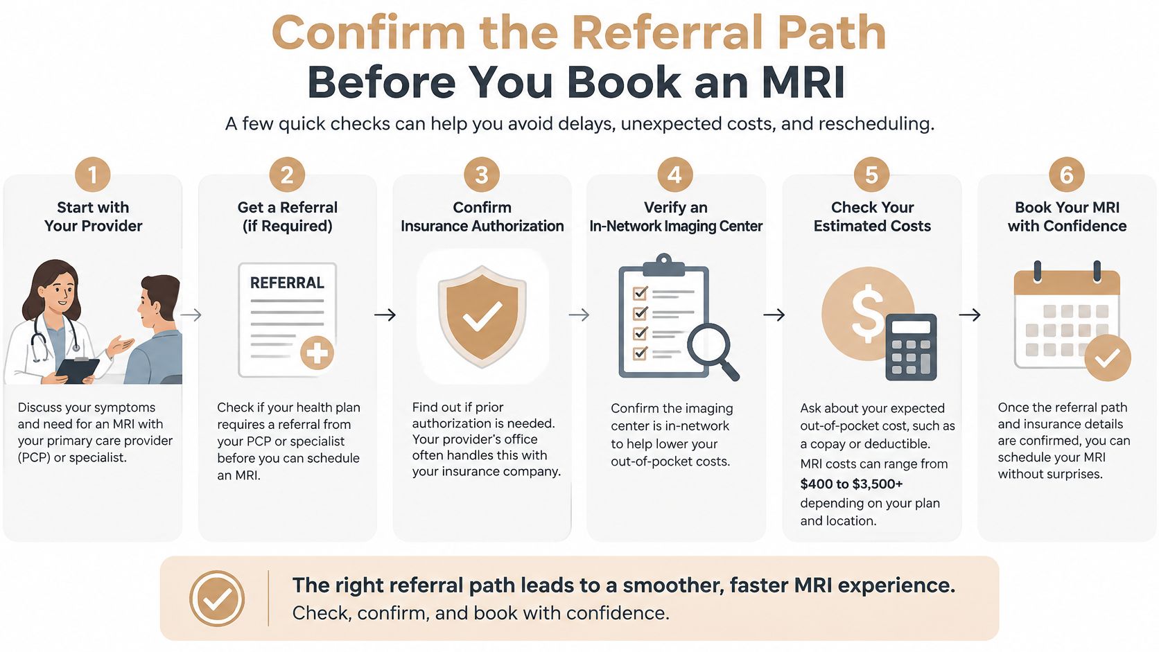
This stepwise approach matches wider guidance. Routine MRI generally isn't recommended at the first visit for uncomplicated low back pain, whether or not sciatica is present, and imaging should be considered when the result is likely to change management, as stated in the NICE guideline recommendations for low back pain imaging. Even a specialist referral doesn't automatically mean you need a scan.
Aspen Falls Wellness starts this pathway with a thorough examination and an X-ray or MRI referral when indicated. Care options include non-surgical Spinal Decompression with the DRX 9000 or MLS Laser Therapy. When your needs fall outside in-house scope, you'll be referred to external specialists.
One question decides the next move: would the image change what you do from here? With any emergency sign, go to emergency care now. With pain but no red flags, book the exam in Salt Lake City or Sandy and put the review date on your calendar today.

Stay updated with the latest chiropractic news and exclusive offers.