Compressed spine pain explained: causes, red-flag symptoms, when imaging helps, and conservative care options such as decompression and rehab.
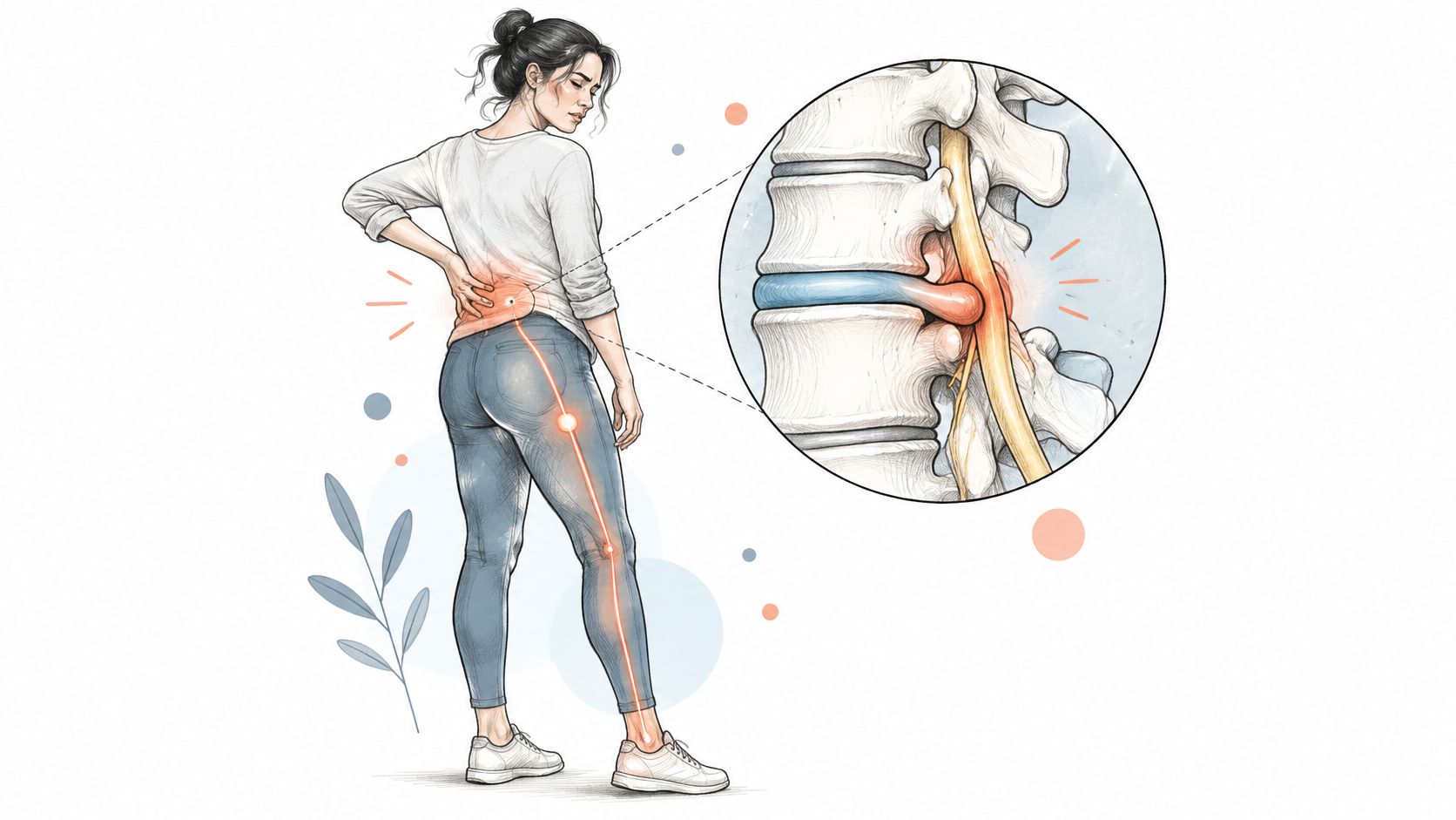
You wake with a stiff lower back, expecting it to loosen up after breakfast. Then pain runs down one leg as you put on your shoes, and you wonder whether this is ordinary soreness or a nerve problem. That difference matters more than the label on a scan, because the safest next step follows your symptoms, nerve function, and response to care.
Compressed spine pain is a symptom pattern, not a single diagnosis, describing pain that may occur when a disc, joint, ligament, or narrowed spinal passage irritates or squeezes a nerve.
Common symptom patterns and what they may suggest
| Symptom pattern | Where it is felt | What it may suggest |
|---|---|---|
| Localised low back ache, stiff with movement | Lower back only | Muscle or joint strain; disc irritation is possible |
| Sciatica-type pain | Buttock down the back of the leg, often below the knee | Nerve root irritation from a disc or narrowed opening |
| Numbness or tingling | Foot, toes, or leg | Sensory nerve root involvement |
| Leg weakness or foot drop | Leg or foot muscles | Motor nerve involvement; needs a clinical check |
| Pain worse with sitting or bending forward | Back and leg | Often disc-related; posture changes the symptoms |
| Pain worse standing and walking, eased by sitting | Lower back and legs | A narrowed spinal canal, or stenosis, pattern |
| Neck pain with arm pain or finger tingling | Neck, shoulder, arm, and hand | Possible cervical nerve root compression |
These are typical presentations, not diagnoses. Pain below the knee raises suspicion for nerve involvement, but its location alone doesn't establish what's causing it. Likewise, tingling in a foot can originate outside the spine, so an examination needs to consider other explanations.
A sore back can make movement uncomfortable without interfering with nerve signals. Nerve involvement becomes more concerning when pain travels into a limb alongside altered sensation, reduced strength, or reflex changes.
For example, avoiding stairs because they hurt isn't the same as being unable to lift the front of your foot. Tell the examiner whether a task is painful, physically difficult, or both.
The Global Burden of Disease 2017 study estimated age-standardized low-back-pain prevalence at about 7.5% in 2017, with 577.0 million people affected, an increase in absolute cases driven by population growth and aging rather than a fall in importance. Those figures describe low back pain overall, not confirmed spinal compression.
Not every painful back has an identifiable structural cause. Symptoms can suggest nerve involvement, but a clinical examination is needed to assess it, sometimes with further testing. Track pain severity and everyday function alongside any imaging findings.
How you load your back can affect symptoms, even when the underlying structure does not change. Prolonged sitting, repeated lifting with a rounded back, and deconditioning can make movement harder to manage. Breaking up sitting, building strength gradually, and adjusting activities that provoke symptoms may help you stay active.
The spine is a stacked column with a central canal and openings between the vertebrae. The spinal cord passes through the upper part of the canal, while nerve roots continue below it and exit through the openings.
The illustration shows several structural ways the available space can change.
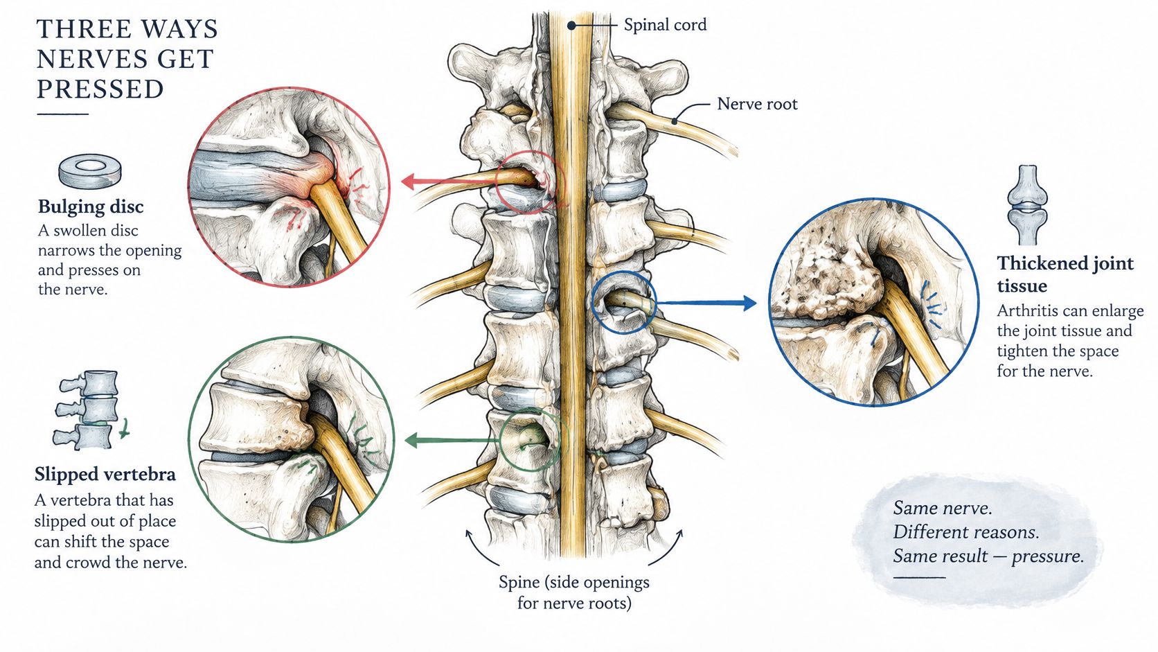
Disc herniation. A disc has a tougher outer layer around a softer center. When disc material pushes outward, it can contact a nearby nerve root. Local inflammation can also irritate the nerve, even without direct pressure.
Foraminal narrowing. The foramen is the opening where a nerve root exits the spine. Disc-height loss or enlargement of nearby joints can shrink this opening and crowd the nerve root.
Spinal stenosis. Narrowing of the central canal can affect nerves traveling through it. This can contribute to symptoms that appear or worsen with standing and walking.
A 2024 population-based cohort study published in Neuroradiology found that 141 of 644 participants, or 21.9%, had at least one lumbar foraminal stenosis. This describes the study population; symptoms and examination findings help determine what the narrowing means for an individual.
Age-related changes may be present without causing symptoms. Changes in posture or activity cannot reverse every structural change, but they can help make movement more manageable. Adjusting repeated lifting that provokes symptoms and discussing body-weight concerns without blame may also support activity.
The practical goal is to reduce irritation and improve movement, even when the spine's anatomy does not return to an earlier appearance. How imaging findings affect care depends on the full clinical picture, including symptoms and response to treatment.
A few symptoms change the decision from waiting to acting the same day. Don't use home treatment or a routine appointment as the only response to the following warning signs.
NICE guidance on low back pain and sciatica excludes progressive neurological deficit and cauda equina syndrome from routine sciatica pathways. Suspected cauda equina syndrome shouldn't be managed with routine conservative care alone.
Use this visual reminder to recognize symptoms that shouldn't wait.
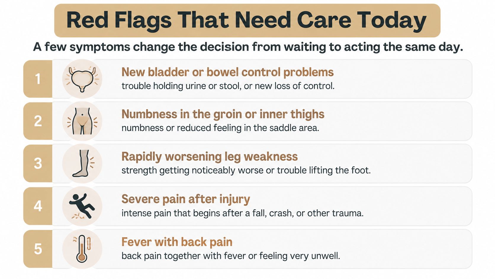
For bladder changes, saddle numbness, or rapidly progressing weakness, go to an emergency department immediately. Other warning signs need same-day medical assessment through urgent care or an emergency department, depending on severity.
Bring a written list of symptoms, when they started, and how they've changed. If weakness makes walking or driving unsafe, arrange help rather than driving yourself.
Our guide to low back pain red flags offers additional context, but reading shouldn't delay urgent assessment. Most nerve-related pain isn't an emergency. These signs are the exception.
Suppose an MRI shows a disc bulge on one side, while your symptoms and examination point to a nerve on the other. That mismatch matters: a scan finding is most useful when it fits the symptoms and could change the next treatment decision.
| Situation | Imaging usually needed? | Typical next step |
|---|---|---|
| Acute low back pain with no red flags | No routine MRI or CT | Monitor, stay active, and reassess |
| Symptoms improving with clinician-guided care | No | Continue care and track progress |
| Radiating leg pain persisting beyond about six weeks | Often, when the result would change treatment | Clinical review, then MRI if a procedure or specialist plan is being considered |
| No improvement after clinician-directed care | Yes, when needed to guide the next treatment decision | Clinical reassessment and possible specialist referral |
| Red flags present, as described in Red Flags That Need Care Today | Urgent imaging may be needed | Same-day emergency evaluation |
| Suspected fracture, infection, cancer, or major trauma | Yes, with the imaging type selected medically | Urgent medical evaluation |
The Healthy Blue Kansas spinal imaging policy supports selective imaging for uncomplicated acute back pain or radiculopathy, including reassessment after approximately six weeks of appropriate care when symptoms persist. Early imaging in people without serious pathology hasn't been shown to improve pain, function, or quality of life.
A thorough chiropractic or medical examination helps identify the likely source of symptoms and whether further investigation is necessary. The examiner checks movement, strength, sensation, reflexes, and walking, then considers the history and response to treatment.
Aspen Falls Wellness provides a thorough chiropractic evaluation to identify the root cause of a patient's issues, followed by a customized treatment plan based on the diagnosis. Dr. Jeremy Kaier specializes in treating herniated discs, stenosis, and sciatica. Diagnostic uncertainty or concerning findings can still call for medical referral before treatment begins.
A disc bulge becomes more relevant when its side and location match the symptoms, nerve distribution, and examination findings. A mismatch may mean the scan finding does not explain the pain.
Treatment decisions follow the full clinical picture and how symptoms respond to care. Manual treatment or a device cannot be assumed to mechanically remove every bulge. A good plan targets movement and symptom control.
A symptom pattern can suggest nerve irritation, but it does not confirm the cause. Treatment choices should match the problem being addressed and change according to how symptoms and function respond. For chronic primary low back pain, structured exercise and rehabilitation have stronger evidence than claims that a passive treatment permanently uncompresses a nerve.
| Option | Best-supported use | Evidence strength |
|---|---|---|
| Structured exercise and rehabilitation | Pain and function in chronic primary low back pain | Moderate certainty from a systematic review |
| Spinal decompression, including DRX9000 or similar traction devices | A selectively considered option for back or radiating pain | Limited; no demonstrated advantage over conventional traction |
| Chiropractic adjustment | Joint motion restriction and pain relief within combined care | Standalone nerve-root benefit is not established by the cited evidence |
| Massage therapy | Muscle guarding and soft-tissue tension | Comfort-focused; direct nerve-root benefit is not established |
| MLS laser therapy | An adjunct intended for pain relief in soft-tissue and inflammatory conditions | Not evaluated by the reviews cited here |
| Acupuncture | Pain modulation alongside other care | Nerve-root-specific benefit remains uncertain |
A 2023 systematic review informing a World Health Organization guideline analyzed 13 trials involving 1,362 adults with chronic primary low back pain. It found moderate-certainty evidence that structured exercise improved immediate pain and physical function. Aerobic, Pilates, and motor-control exercise were associated with pain reduction; mixed programs and Pilates were associated with functional improvement. Certainty for individual exercise types was lower.
A 2023 meta-analysis of traction approaches found no statistically significant superiority of spinal decompression over conventional mechanical traction for pain or disability. It also found no meaningful difference between high-force and low-force traction. The table summarizes this finding: a device name or setting does not establish better results.
NICE recommendations on decompression for sciatica concern surgical decompression, considered when nonsurgical treatment has not improved pain or function and imaging findings match the sciatic symptoms. That recommendation concerns surgical decompression, not traction devices such as DRX9000.
Manual care and massage may make movement more comfortable. That comfort alone does not show whether nerve irritation has settled. Laser therapy and acupuncture can also be considered for specific symptom goals, with expectations guided by the evidence for each option.
A 2025 systematic review of nonsurgical interventions for acute and subacute sciatica rated the overall evidence very low confidence because of bias and imprecision. Combined non-pharmacological care may improve low-back-pain outcomes compared with a single intervention, but the same advantage cannot be assumed for leg pain.
A useful treatment earns its place by helping you walk, sleep, work, or exercise more comfortably. Agree on a measurable goal and a point to reassess before continuing any treatment indefinitely. For more context, read this guide to managing nerve pain.
Break up sitting every 30 minutes with a brief walk or standing break. If sitting brings on leg symptoms sooner, change position earlier. Keep movement comfortable and build walking distance gradually, stopping before symptoms flare.
For lifting, bend through your hips and knees, keep the load close, and reduce repeated rounded-back lifting if it aggravates your pain. At night, try side sleeping with a pillow between your knees or back sleeping with one under them. Keep the position that feels more comfortable.
Notice how symptoms respond to movement. If a stretch or exercise sends pain farther down your leg, stop and record what triggered the change so your clinician can adjust the plan. During a flare, brief rest may help; resume tolerable movement as you are able.
Use these steps alongside your prescribed home exercises. On a difficult day, you can skip an exercise that clearly worsens limb symptoms and ask your clinician how to modify it.
If your sleep surface seems to aggravate discomfort, Slone Brothers Furniture's guide to mattress firmness for back pain relief offers practical shopping context. Choose a mattress for comfort; firmness does not treat nerve compression.
For new or worsening warning signs, follow the guidance in Red Flags That Need Care Today. Self-care can support your routine between visits, while persistent or changing symptoms may call for a clinical review.
A back or leg symptom can change before its cause is clear. If symptoms follow a nerve pattern and persist, book an evaluation so a clinician can check strength, sensation, and reflexes and decide whether further testing could change your care.
Recovery timeline with escalation triggers
| Timeframe | Typical sign to review | Seek reassessment if |
|---|---|---|
| First 1 to 2 weeks | Pain begins easing with brief rest and gentle movement | Leg weakness or bladder changes appear |
| Weeks 2 to 6 | Walking tolerance and sleep improve | Pain stays unchanged or gets worse |
| Weeks 6 to 12 | Radiating pain eases and function continues improving | There is no functional gain after structured care |
| Beyond 12 weeks | Function remains stable with fewer flare-ups | Symptoms plateau or recur, prompting discussion of specialist review or imaging |
These are review windows for checking progress. A new or worsening neurological symptom needs attention whenever it appears.
A short record can help you and your clinician spot meaningful changes. Note:
Mood can also provide context alongside sleep and activity, since ongoing pain affects daily well-being. If a visual diary suits you, track moods with Life Calendar, while keeping physical symptoms in a separate log.
At Aspen Falls Wellness, patients typically begin to experience back pain relief within weeks. Progress is evaluated with each adjustment, and care is adjusted based on how the patient is responding. Worsening symptoms may call for a fresh assessment and a change in approach.
Care may include customized chiropractic adjustments, DRX9000 non-surgical spinal decompression, MLS® Laser Therapy, massage therapy, acupuncture, or rehabilitation exercise, depending on the diagnosis and treatment plan. Aspen Falls Wellness also offers SoftWave therapy, nutrition, mobility therapy, golf movement screening, and car accident treatment.
If your needs fall outside the clinic's scope, referral to an outside specialist may be appropriate. A useful evaluation should clarify the working diagnosis, set realistic goals, and establish what happens if symptoms do not improve.
To arrange an evaluation at Aspen Falls Wellness in Salt Lake City or Sandy, visit Aspen Falls Wellness and bring your symptom log to discuss the next step.

Stay updated with the latest chiropractic news and exclusive offers.