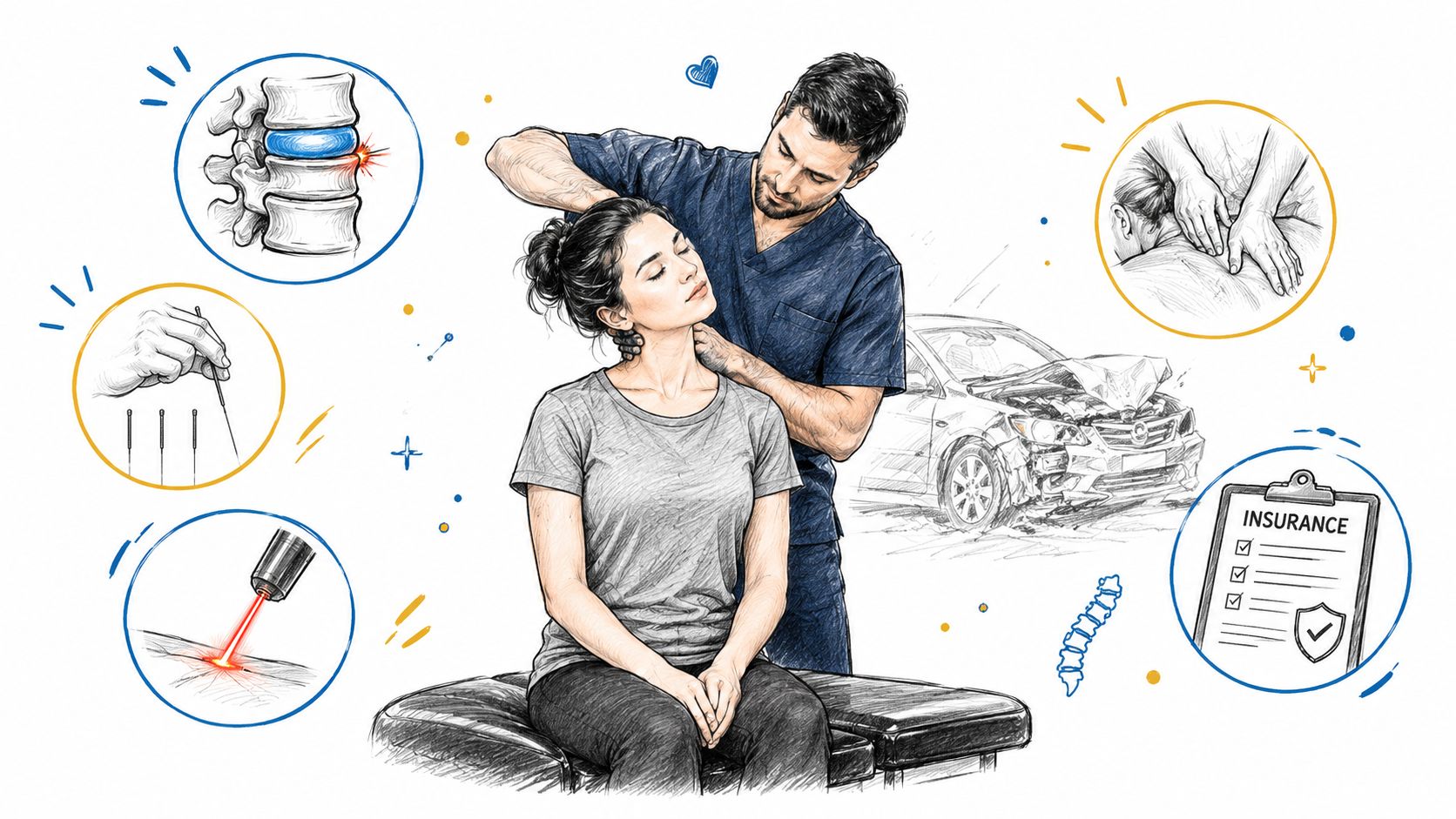
Understand disc herniation and sciatica causes, symptoms, and evidence-based non-surgical treatments like spinal decompression at Aspen Falls Wellness in Utah.
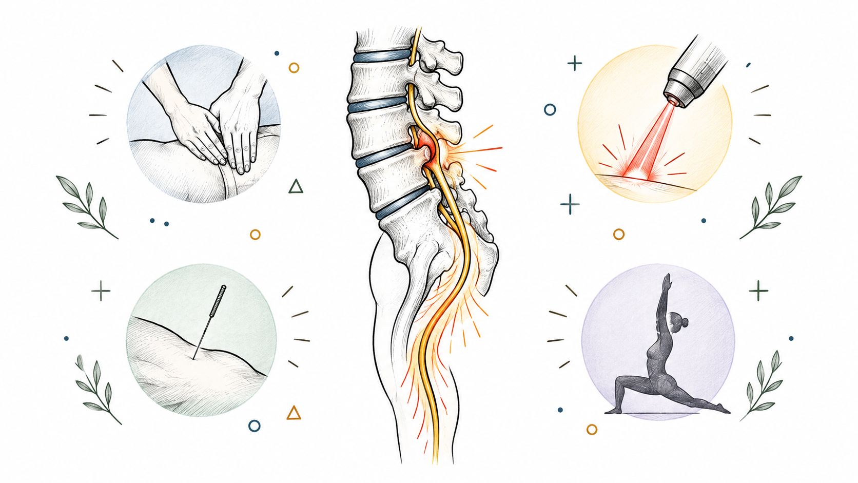
Approximately 85% to 90% of sciatica cases involve lumbar disc herniation that compresses or irritates a nerve root, and conservative, non-surgical care helps approximately 80% of patients improve within four to six weeks.
You may be sitting in your car, turning in bed, or reaching for something on a low shelf when a sharp pain travels from your lower back into your buttock and down your leg. The sensation can feel alarming, especially when numbness, tingling, or weakness joins the pain. You may also have an MRI report describing a “bulging” or “degenerative” disc and wonder whether that image explains everything.
Often, it doesn't. A disc finding matters when it matches your symptoms and neurological examination. The most useful question isn't, "What does the MRI show?" It's, "Do the symptom pattern, examination, and imaging point to the same irritated nerve?"
A patient may describe the pain as starting in the lower back, crossing one side of the buttock, and continuing down the back or side of the leg. Sitting makes it worse. Coughing or bending may intensify the jolt. This pattern differs from ordinary muscle soreness because the pain follows a nerve-root distribution.
The spine's discs act as cushions between the vertebrae. Each one has a firm outer ring, called the annulus fibrosus, and a softer inner center, called the nucleus pulposus. If the outer layer weakens or tears, the inner material can move outward. That event is called disc herniation.
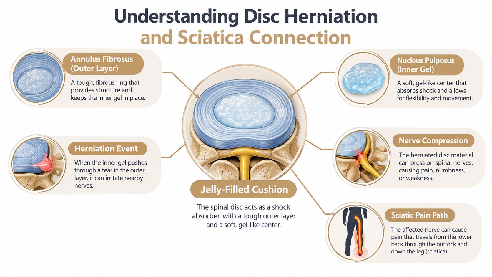
A lumbar herniation can compress or inflame a nearby nerve root. Those nerve roots contribute to the sciatic nerve, so irritation can create pain, tingling, altered sensation, or weakness farther down the leg. The problem isn't necessarily that the entire sciatic nerve is damaged. More often, a nerve root is being affected near the lower spine.
Evidence summarized in a clinical review indicates that a herniated disc causes lumbar nerve-root compression or inflammation in approximately 85% to 90% of sciatica cases (clinical review of sciatica). Sciatica itself means radiating pain from the lower back into the buttock and leg along a lumbosacral nerve-root distribution. It isn't interchangeable with nonspecific low-back pain.
Symptoms can also arise from inflammation around the nerve, not only direct physical pressure. That distinction helps explain why someone may have severe leg pain even when the disc protrusion appears modest, while another person may have a more noticeable MRI abnormality with little or no pain.
Practical rule: A disc image becomes clinically meaningful when it agrees with the side, route, sensory changes, strength findings, and reflex changes found during the examination.
Foot mechanics can affect how you load the legs and pelvis during walking, although arch support isn't a treatment for nerve compression. If foot discomfort contributes to altered movement, patients may find it useful to review general sciatica arch support advice alongside a proper spinal assessment.
You wake up with sharp pain shooting from the low back into the leg, and the first thought is often, “Do I need an MRI right now?” In many cases, the better first step is a careful exam. Spinal scans can show disc bulges and wear that have nothing to do with your current pain, so the primary task is matching the story, the examination, and the image.
For uncomplicated acute sciatica, routine MRI during the first four to six weeks is generally discouraged when there are no red flags (acute lumbar disc herniation evaluation). That waiting period is not dismissal. It is a structured observation phase in which a clinician tracks whether the irritated nerve is calming down, staying the same, or losing function.
A useful way to picture the process is as a three-part match. First comes the symptom map. Then the neurological exam. Imaging comes after that, when it can confirm the suspected level and side of nerve-root involvement or change what happens next.
Map the symptoms. Your clinician should ask where the pain starts, how far it travels, whether numbness or tingling follows the same path, and what positions make it worse or better. Pain that runs below the knee or reaches the foot can suggest a different nerve-root pattern than pain limited to the buttock. Delayed onset can also matter, especially after trauma, so a written record of symptom timing may help both medical follow-up and practical matters such as documenting symptoms for a claim in Colorado.
Check neurological function. The examination should include dermatomal sensation, myotomal strength, reflexes, gait, and simple functional movements such as heel or toe walking when appropriate. A straight-leg-raise test can reproduce radiating leg pain, but it does not stand on its own. The question is whether the exam findings line up with the symptoms on the same side and in the same nerve distribution.
Screen for urgent warning signs. Some symptoms need immediate escalation rather than routine follow-up. Urgent warning signs are covered in detail in the final section.
Use imaging when it can change management. MRI becomes more useful when symptoms continue beyond approximately six weeks, when an injection or surgery is being considered, or when severe or progressive neurological findings appear. At that point, the scan is most helpful if it matches the clinical picture instead of showing age-related change.
That concordance matters. A disc finding is more likely to be meaningful when it fits the side of the pain, the route down the leg, the numb area, the weak muscle group, and any reflex change. If those pieces do not fit together, the image may be incidental rather than explanatory. Patients who want a plain-language overview of these symptom patterns can review how to know if you have a herniated disc.
Follow-up imaging can also be confusing. A scan may still look abnormal while the person is improving, or it may show partial settling of the herniation over time. A major review found complete or partial resolution in 64 of 84 documented herniations, or 76%, after one year (history and natural course of lumbar disc herniation). That is why staged decision-making works better than treating every MRI finding as an emergency.
You wake up with the same leg pain again, and the hardest question is not whether a disc is visible on MRI. It is whether the scan, your symptoms, and your examination all point to the same irritated nerve, and whether that nerve is recovering or falling behind.
For patients whose findings line up clearly, meaning the MRI-confirmed herniation matches the side of pain, the nerve pattern, and the neurological exam, early discectomy can relieve leg pain faster by removing material that is pressing on the nerve. Faster relief, though, is not the same as a better long-term result. In a randomized trial involving 501 patients with at least six weeks of sciatica, both early surgery and prolonged conservative care led to substantial improvement, with no meaningful difference in clinical outcomes after one and two years.
The first phase of care is active, not passive. It usually includes education, activity changes, medication when appropriate, rehabilitation exercise, and close follow-up. The goal is to keep you functioning while the irritated nerve has time to settle, and to watch for signs that recovery is actually happening.
That progress is measured in ordinary things. Walking a little farther. Sitting a little longer. Sleeping with fewer wake-ups. Feeling pain move out of the calf and toward the back or buttock instead of spreading farther down the leg.
Over the next several weeks, the clinician is asking a very specific question. Are symptoms improving in a pattern that fits nerve recovery, or is weakness, numbness, or function getting worse despite treatment? If the pain remains disabling or neurological function declines, the decision changes. An injection may help some patients get short-term relief, and surgery becomes more reasonable when persistent radiculopathy continues to limit daily life or weakness progresses.
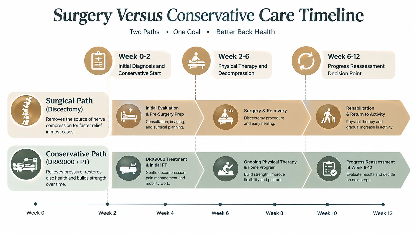
Natural disc regression helps explain why nonoperative care often succeeds. A clinical review reports that symptoms commonly resolve within four to six weeks and that conservative treatment is effective for approximately 80% of patients during that period.
The timeline works best as a staged decision process. If symptoms, examination findings, and imaging continue to match and recovery is underway, conservative care often makes sense. If they match and the nerve is not recovering, faster intervention may make more sense.
You wake up with leg pain that shoots below the knee, and the MRI report lists several disc changes. The next question is not which treatment sounds strongest. It is which treatment fits the problem your clinician can confirm. Non-surgical care works best when symptoms, examination findings, and imaging point to the same irritated nerve, and each treatment is chosen for a clear job.
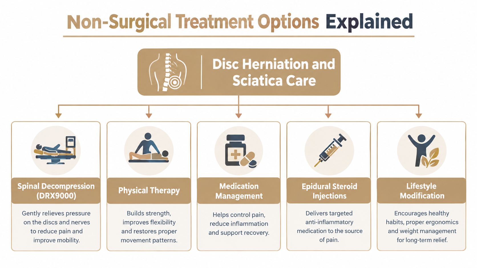
Spinal decompression with the DRX 9000 uses controlled positioning and traction to change how load passes through selected spinal segments. It works more like adjusting pressure on a sore hinge than forcing a disc back into place. Before using it, a clinician should check whether your pain pattern, nerve findings, and tolerance make this type of loading reasonable. Patients who want more background can read about spinal decompression therapy before deciding whether it belongs in a real treatment plan.
Chiropractic adjustment, realignment, and mobility therapy aim to improve motion in stiff areas and reduce the protective bracing that often builds around back and leg pain. That matters because guarding can make every step feel threatening, even when the nerve itself is starting to settle. The approach should match the person in front of the clinician, especially if radiating pain, numbness, or weakness is present.
SoftWave Therapy and MLS Laser Therapy are device-based treatments used to support local tissue care. Their role is narrower than many patients expect. They may address pain-sensitive tissues around the area, but they do not replace the work of confirming whether the main driver is true nerve root compression, local inflammation, or muscular guarding.
Massage Therapy can reduce muscle tension in the low back, hip, and leg. It does not remove a herniation, but it may lower enough guarding to help someone walk more normally or tolerate exercise again.
Acupuncture may help calm pain signaling for some patients. Muscle Stimulation may have a place when the examination shows poor muscle activation or weakness, especially if it supports a broader rehabilitation plan rather than standing alone.
Active rehabilitation usually carries the most lasting value. Personal Exercise Plans, rehabilitation exercise, posture retraining, and mobility work help rebuild tolerance for bending, walking, lifting, and returning to daily tasks. The program should change as symptoms change. If leg pain centralizes, strength improves, and nerve tension signs ease, progression makes sense. If weakness spreads or the pain pattern stops matching the original diagnosis, the plan needs reassessment, not just more visits.
Medication decisions and epidural steroid injections belong with qualified medical professionals. A review of injection approaches suggests some injections may offer earlier relief, while regenerative options are still developing rather than serving as a standard answer for everyone. Patients exploring options such as find sciatica relief with stem cells should ask how the treatment matches the diagnosis, what risks it carries, and what alternatives exist.
A clinic may combine these services selectively. At Aspen Falls Wellness, that can include chiropractic care, Spinal Decompression with the DRX 9000, MLS Laser Therapy, massage, acupuncture, mobility therapy, nutrition counseling, and rehabilitation exercise. The goal is not to stack treatments. The goal is to choose the few that match the stage of recovery you are in.
A coordinated plan works best when everyone is solving the same problem. In disc herniation and sciatica, that means matching the person's symptoms, exam findings, and imaging before stacking treatments. An MRI can show age-related disc changes that are present but not driving the pain. A team adds value when it keeps asking a simple clinical question: does this treatment fit the irritated nerve pattern in front of us, and is function improving?
That shared reasoning also helps explain why two people with the same scan may need very different care. One may need help calming muscle guarding just to walk normally again. Another may be ready for progressive loading because leg pain is easing and strength is stable. A third needs referral because weakness is spreading or the exam no longer lines up with a routine recovery pattern.
A multidisciplinary plan should assign each treatment a job. Manual care may help with painful stiffness or movement avoidance. Decompression may be used as a controlled loading strategy. Laser or soft tissue treatment may reduce local irritability enough for exercise to become tolerable. These options are not interchangeable, and they should not all start at once unless there is a clear reason.
Home practice carries the plan into real life. Rehabilitation exercise and mobility work help you test whether the nerve is becoming less sensitive during sitting, walking, bending, and daily tasks. Nutrition counseling may support recovery habits. Golf Movement Screening can be useful when return-to-play mechanics matter to an active golfer.

Combined care can make sense when each part has a defined purpose and the patient is reassessed regularly. For example, some interventional approaches have been studied alongside other pain procedures, but that does not mean every person with sciatica needs a layered treatment package early.
The team's value comes from using examination findings and progress measurements to decide what belongs in your plan. Useful markers are concrete: walking tolerance, sitting tolerance, sleep, strength, gait, and whether the pain pattern is centralizing or spreading. Patients who want an overview of this care model can read about multidisciplinary pain management.
The safest treatment decision follows a sequence rather than a popularity contest. Start with urgency, continue with a defined conservative trial when appropriate, and escalate when function or neurological status calls for it.
Urinary retention, loss of bowel or bladder control, saddle anesthesia, and progressive motor weakness require immediate medical evaluation. These findings can indicate serious nerve compromise and shouldn't be managed through routine appointments alone.
Pain scores matter, but function often gives a clearer signal. Track whether you can walk farther, sit longer, sleep more consistently, put on shoes, or complete prescribed exercises. A person whose pain remains present but whose walking and strength improve may be recovering in a meaningful way.
A structured conservative trial can include activity modification, education, rehabilitation exercise, mobility therapy, and appropriately selected symptom-relief treatment. A review of persistent sciatica identifies six to eight weeks as a common period for reassessment and possible escalation, while severe pain and disability often improve within two to four weeks (review of persistent sciatica).
If progress has stalled, an epidural injection may be discussed for symptom relief, or a surgical consultation may be appropriate for persistent disabling radiculopathy. A striking MRI alone shouldn't make that decision. The strongest case for escalation comes from concordance between the symptom route, neurological findings, functional limitation, and relevant imaging.
This approach avoids two opposite errors: rushing into a procedure because an image looks dramatic, or continuing passive treatment indefinitely while weakness progresses.
Patients with disc-related sciatica don't develop a surgical emergency, but certain symptoms require prompt action. Don't wait for a routine follow-up if you develop urinary retention, loss of bowel or bladder control, or numbness in the saddle region around the groin and inner thighs.
Cauda equina syndrome can involve severe compression of the nerves responsible for bladder, bowel, and pelvic sensation. It requires immediate medical assessment. Bilateral leg symptoms or rapidly changing neurological function make the situation even more urgent.
Substantial motor weakness, including strength measured at grade 3 or less, warrants prompt specialist evaluation. In practical terms, you may be unable to lift the foot against gravity, maintain knee extension, or perform a movement that was previously easy.
Progressive weakness means strength is deteriorating over days rather than gradually returning. Spreading numbness, a worsening gait, or increasing difficulty lifting the front of the foot also deserves urgent attention. Persistent nerve-root pain associated with foraminal stenosis may require specialist review when decompression and rehabilitation aren't producing adequate progress.
Seek immediate care for: urinary retention, bowel or bladder loss, saddle anesthesia, rapidly worsening weakness, or severe neurological decline.
Aspen Falls Wellness can evaluate symptoms, document neurological findings, provide non-surgical options when appropriate, and coordinate referral when the presentation falls outside conservative care. Visit Aspen Falls Wellness to discuss an examination-led plan for disc herniation and sciatica, including chiropractic care, decompression, rehabilitation exercise, and follow-up based on your progress.

Stay updated with the latest chiropractic news and exclusive offers.