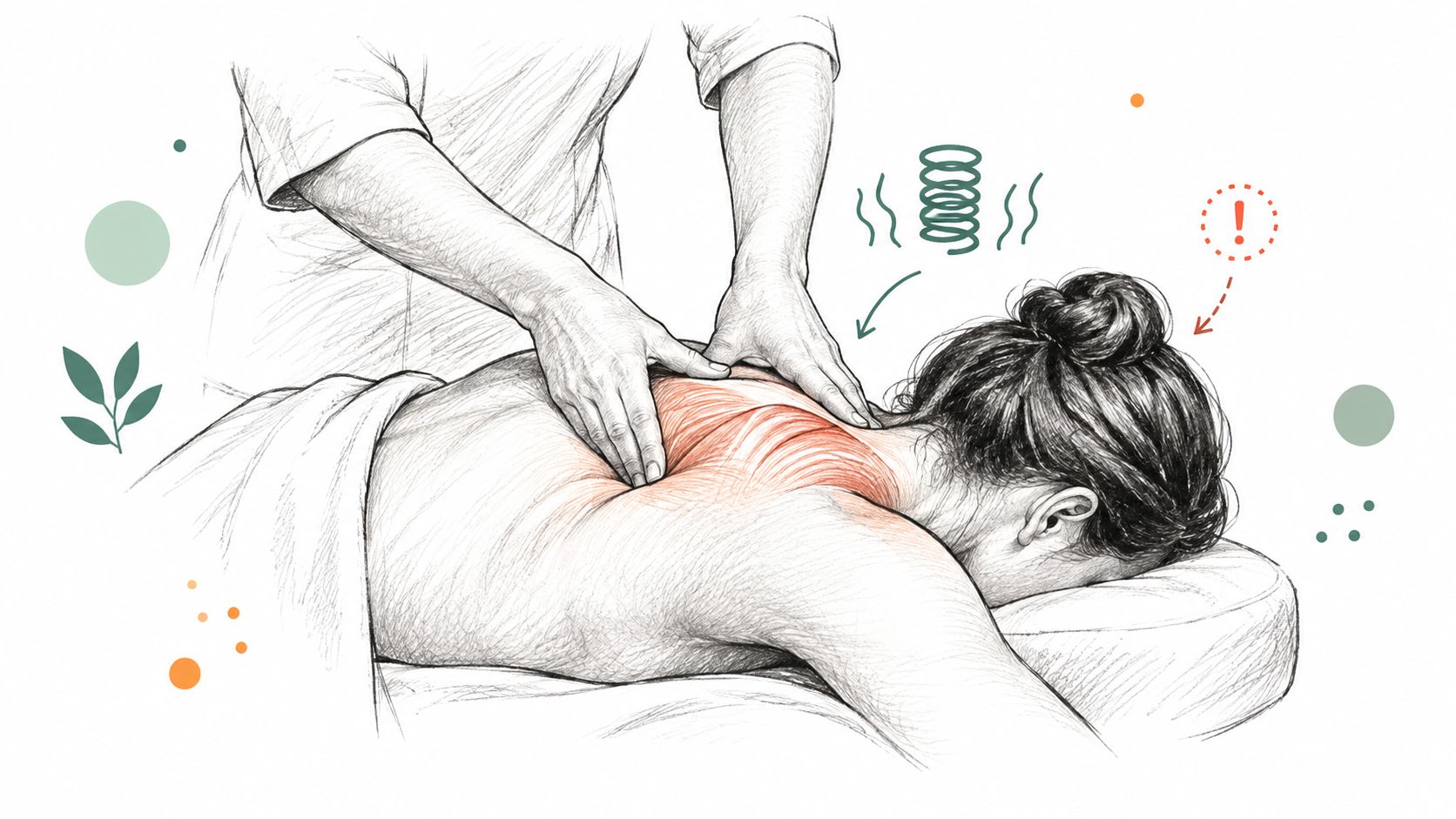
Confused about mri vs xray for back pain? Learn what each scan shows, when it's needed, and how our experts determine your best path forward.
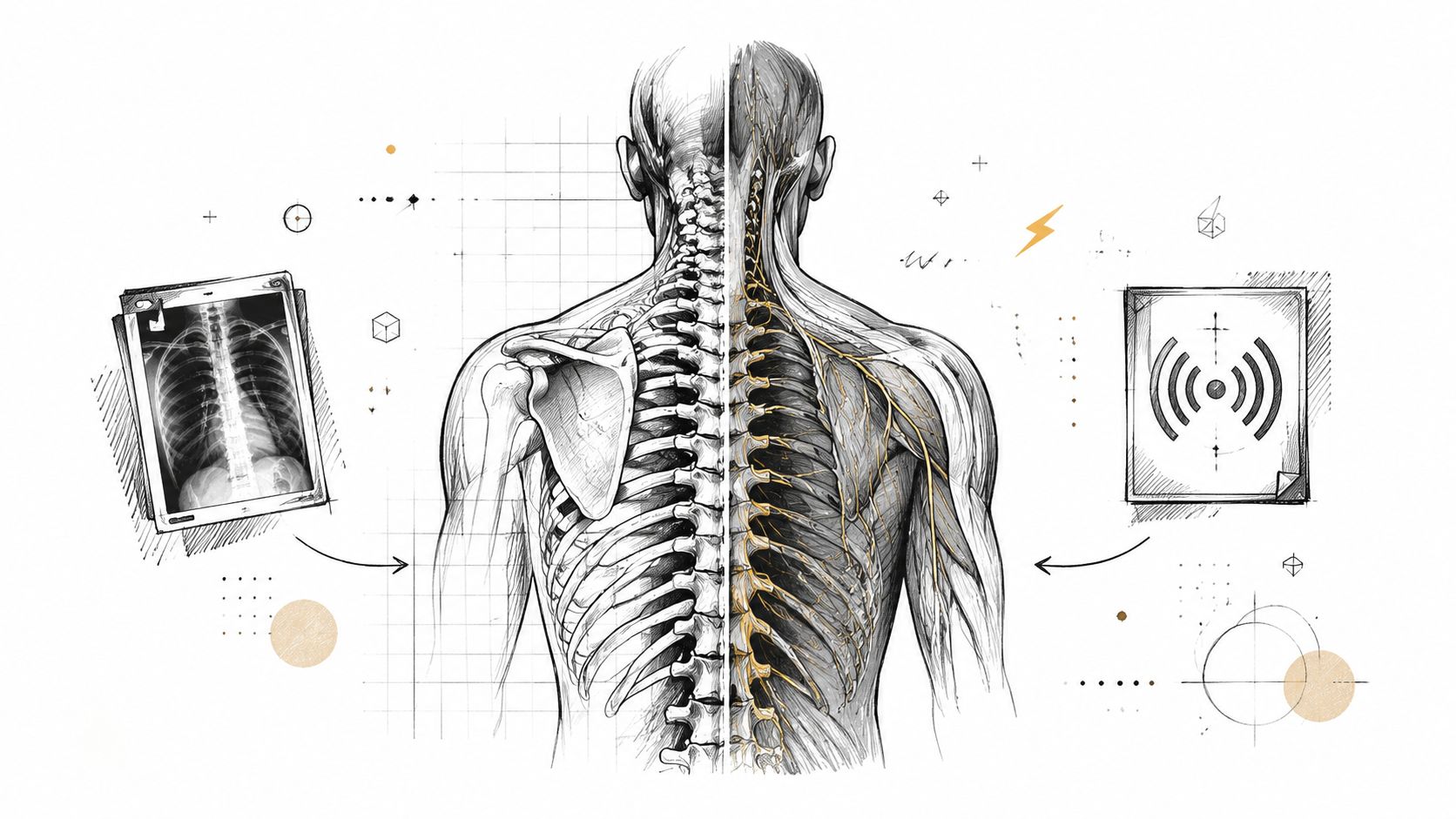



The most common advice about back pain is also the least useful: get an MRI as soon as possible. In real practice, that's often the wrong first question. The better question is whether imaging is needed at all, because for many cases of uncomplicated low back pain, a thorough exam and a short period of conservative care come before any scan.
That approach isn't a delay tactic, it's evidence-based triage. When imaging is needed, MRI vs Xray for back pain isn't a contest between “better” and “worse.” It's a decision about what problem the clinician is trying to rule in or rule out, and whether the symptoms justify a scan in the first place.
| Test | Best at showing | Main limitation | Common use case |
|---|---|---|---|
| X-ray | Bone, alignment, fractures, degenerative change | Limited soft-tissue detail | Trauma, fracture concern, structural bone questions |
| MRI | Discs, nerve roots, spinal cord, infection, malignancy concerns | More detailed than needed for many simple pain cases | Radicular symptoms, neurologic compromise, soft-tissue pathology |
Most back pain does not need immediate imaging. That is not because the pain is minor. It is because routine imaging for uncomplicated low-back pain has repeatedly shown no clinical benefit, and a review of randomized trials found no meaningful difference between routine lumbar imaging and usual care in pain, function, quality of life, or overall patient-rated improvement (PMC review).
A careful physical exam comes first because symptoms alone do not tell the full story. At Aspen Falls Wellness, that exam helps sort out everyday mechanical pain from patterns that suggest something more urgent, and that is where a clinician decides whether imaging will clarify the problem or just add another result to interpret. For patients trying to understand their own low-back symptoms, a plain-language overview such as this guide to lower back pain can also help frame the discussion before testing is ordered.
Imaging becomes more appropriate when the story includes red flags, not just discomfort. Those include cancer risk, cauda equina symptoms, severe neurologic deficit, significant trauma, suspected fracture, fever, unexplained weight loss, osteoporosis with new severe pain, progressive leg weakness, saddle numbness, or new bladder or bowel loss (Gale Care).
Practical rule: severe pain by itself does not automatically mean MRI. The exam matters more than the intensity of the ache.
For pain that has only been present a short time, many guidelines discourage routine imaging because early scans do not improve outcomes in uncomplicated cases (AAFP). A patient can feel miserable and still not need a scan on day one. That is also why a clinician may focus first on movement testing, neurologic screening, and a close look at how symptoms behave with posture, walking, bending, or coughing.
The clinical question is not, “Which scan is better?” It is, “Is a scan necessary yet?” When the answer is no, that often reflects careful care rather than delay.
An X-ray is the simpler of the two tests to understand. Think of it as looking at the frame of a house. You can see the structure, alignment, and obvious damage, but you can't inspect the wiring or plumbing. For back pain, that means X-rays are useful when the question is about bone, not soft tissue.

X-rays are most helpful for fractures, spinal alignment, and advanced degenerative change. They can show whether the spine looks out of line, whether there's concern for instability, and whether a bone injury is visible after a fall or motor vehicle collision. In older adults, they also help when a compression fracture is on the differential, especially when osteoporosis is part of the story (Camelback Spine Care).
That's why an X-ray often comes first when the concern is structural and bony. It answers a narrow but important question quickly: is the skeleton intact?
X-rays are poor at showing discs, nerve roots, muscles, ligaments, and the spinal cord. They can't tell you much about soft-tissue inflammation or nerve compression, and they're not the best tool for explaining radiating leg pain or numbness (AMA Journal of Ethics).
If a patient's pain pattern suggests nerve involvement, a normal X-ray doesn't rule that out. It only tells you that the bones look acceptable on that study. That distinction matters, because a reassuring X-ray can still leave the actual cause untouched.
For that reason, X-ray is usually a bone-focused test. It's the right choice when the clinician is asking, “Did the spine break, shift, or collapse?” If that's not the question, X-ray often isn't the answer.
An MRI gives a closer look at the structures X-ray cannot show well. Where X-ray is best at bone detail, MRI shows the discs, nerves, spinal cord, and surrounding soft tissues more clearly, so it becomes the better study when symptoms suggest the problem is not limited to the skeleton.

MRI is used when the clinician suspects disc herniation, spinal stenosis, nerve compression, infection, malignancy, or neurologic compromise (AMA Journal of Ethics). It can show the structures that may explain sciatica, numbness, weakness, or pain that follows a nerve pattern into the leg. For patients with symptoms that fit a disc problem, the scan can also guide next steps in disc herniation treatment options by showing whether the disc is affecting nearby nerves.
That is why MRI is the more precise study when the question is not whether the bone is intact, but what is pressing on or irritating the nerve.
More detail is not always better care. In uncomplicated low back pain, routine advanced imaging does not improve outcomes, and evidence reviews have found no meaningful benefit in pain or function from routine radiography, MRI, or CT. A randomized trial summarized by TheNNT found that MRI in patients without pathologic features led to higher cost and more downstream procedures without changing long-term pain, disability, or functional status.
MRI is powerful, but it works best when the clinical question is already narrow.
That is the primary reason it is reserved for the right symptoms. It is not the scan to order first just because it looks more advanced. It is the better choice when the exam and symptom pattern point toward soft tissue or nerve pathology, or when the clinician needs a closer look at a specific concern.
The choice between the two tests becomes clearer when you compare what each one is built to do. X-ray is about bone detail. MRI is about soft tissue detail. Everything else follows from that.
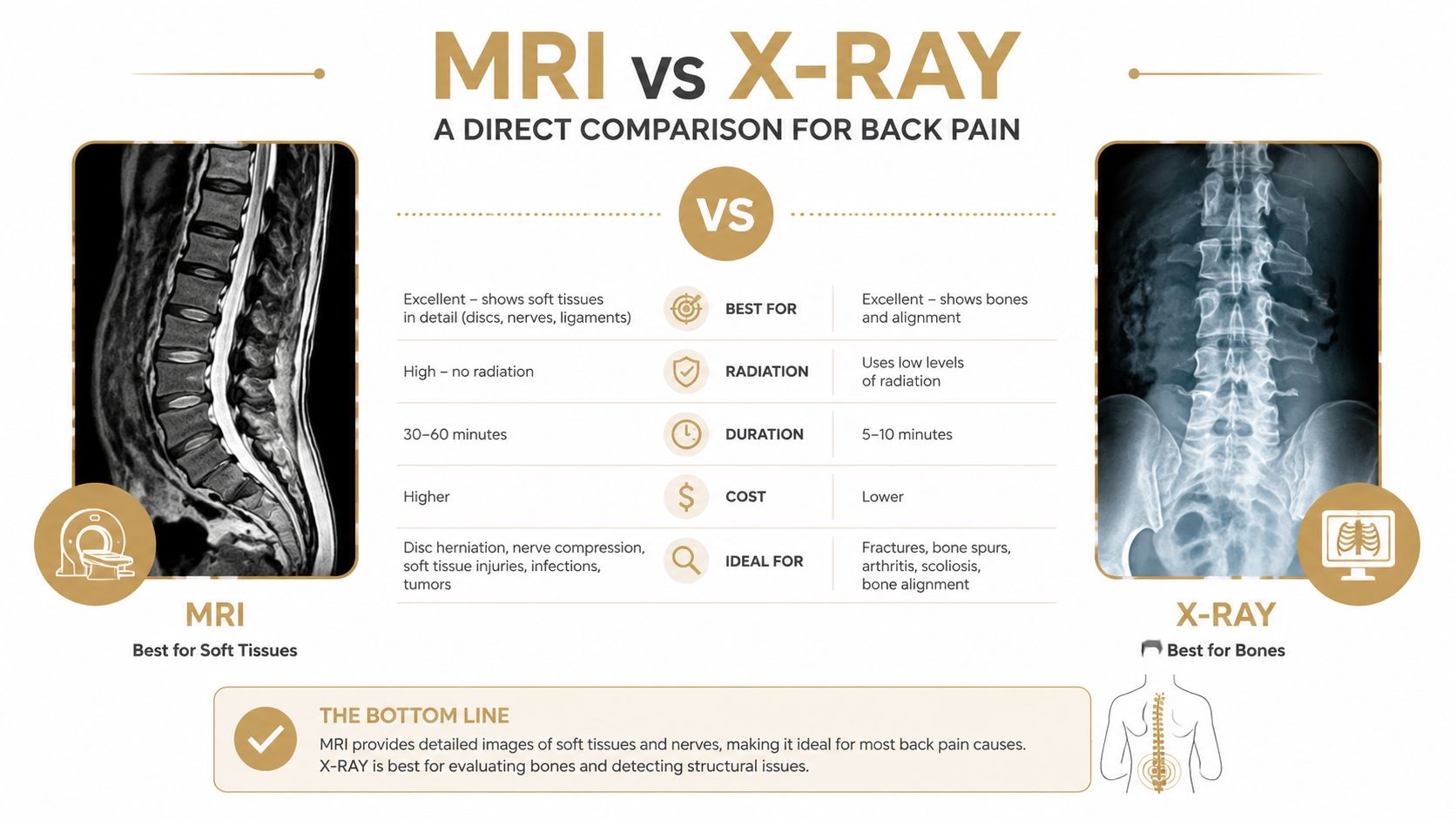
An X-ray uses ionizing radiation, while MRI does not. That matters, especially when repeat imaging might be considered, but the larger issue is diagnostic fit. If the concern is a fracture or alignment problem, X-ray gives the needed answer efficiently. If the concern is a disc, nerve root, infection, or malignancy, MRI is more informative (PMC guideline review).
X-rays are also generally faster and simpler for patients to complete. MRI usually takes longer and can feel more demanding, particularly for people who struggle with tight spaces or staying still. That's not a drawback in every case, but it is part of the decision.
A useful way to think about it is this.
That middle point is the one many people miss. The most important clinical choice often isn't which test to order, it's whether the exam justifies imaging now. For disc-related symptoms, clinicians sometimes move to MRI after a focused exam suggests nerve involvement, and a discussion of conservative options may include disc herniation treatment options when the pattern fits.
A scan can only answer the question it's asked to answer. If the wrong test is ordered, the result may look “normal” and still leave the patient without clarity. If the right test is ordered too early, it can reveal findings that don't explain the pain at all. That's why the best imaging choice starts with symptoms, exam findings, and a clear suspicion about what is being injured.
A car accident changes the equation fast. If the history suggests trauma, a clinician often starts with X-ray because the main question is whether there's a fracture, alignment issue, or other bony injury. That logic also fits older adults with osteoporosis risk, where a compression fracture can happen with relatively minor force.
When the pain shoots down one leg, especially with numbness or weakness, MRI becomes more useful because nerve-root irritation or disc pathology moves to the top of the list. That's the kind of pattern where X-ray can look unrevealing even though the symptoms are very real.
A scan should match the symptom pattern, not just the patient's frustration level.
A different scenario is the athlete with lingering back pain from repetitive loading. That person still needs a physical exam first. If the exam suggests a muscle strain, joint irritation, or movement dysfunction, conservative care may come before imaging. If symptoms persist or neurologic signs appear, the pathway can change.
For persistent, non-specific low back pain, early imaging is often unnecessary. Major guidance says do not image within the first 6 weeks unless red flags are present, and most acute cases improve within that window with conservative care (Gale Care). That's the scenario where patients often expect a scan most, even though the evidence says the scan rarely changes the first step.
A useful resource for people recovering from a spinal injury is Lake City PT fracture recovery help, especially when the concern is how a fracture may affect movement, sitting tolerance, and the next phase of rehab. That kind of support matters because imaging is only part of the pathway, not the whole plan.
The pattern across all four scenarios is the same. The clinician is matching the test to the suspected cause, not ordering a scan just because pain is present.
Unnecessary imaging can create problems that do not help the patient feel better. One of the biggest issues is incidental findings, age-related changes that show up on scans but are not the source of pain. Once they appear on a report, they can trigger worry, extra visits, and sometimes procedures the patient did not need in the first place.
Cost is another concern. In patients without pathologic features, a randomized trial summarized by TheNNT found that MRI led to higher cost and more downstream procedures without improving long-term pain, disability, or functional status. That matters in real practice. A scan can use resources without changing the outcome that matters most, how the person feels and functions.
Guidelines and reviews consistently note that early imaging for uncomplicated low back pain does not improve pain or function, and routine radiography, MRI, or CT is not associated with clinically meaningful benefit (PMC review). Selective imaging is the evidence-based approach. Routine imaging is not.
MRI can also reveal normal wear-and-tear that looks dramatic on paper but has little to do with the current flare. For patients, that can turn a manageable episode into a more confusing one. For clinicians, it can pull the conversation away from movement, rehab, and recovery and toward findings that do not need treatment.
More information is not always more clarity.
A careful exam is often the better first step. It helps separate the cases that need imaging from the ones that are better served by a physical examination, symptom tracking, and conservative care. At Aspen Falls Wellness, that is the point of the workup before any scan is ordered. When disc symptoms or nerve irritation are part of the picture, spinal decompression therapy may fit into the care plan. For patients trying to avoid morning back pain, sleep position and the setup around recovery also matter, and avoid morning back pain can be a useful place to start.
At Aspen Falls Wellness, care begins with a thorough initial examination. We look at movement, posture, neurologic signs, symptom behavior, and the story behind the pain, because imaging only belongs in the plan when the exam points to a clear reason for it. If a scan is needed, it is ordered to answer a specific clinical question, not as a reflex.
A careful exam often comes before any image because back pain does not always come from the same structure. If the findings point to a disc problem, nerve irritation, or a sciatica pattern, the plan may include Spinal Decompression with the DRX 9000, along with targeted chiropractic adjustment, mobility work, and home exercise. If the exam points more toward muscle strain, soft-tissue irritation, or guarding, the pathway may include MLS Laser Therapy, massage therapy, SoftWave Therapy, muscle stimulation, and rehabilitation exercise. The goal is to match care to the actual driver of the pain, not just the label on an image.
That approach also guides care after trauma or when a structural problem is suspected. Imaging can shape the plan, but treatment still has to fit the person in front of us. If disc symptoms or nerve irritation are part of the picture, spinal decompression therapy may be part of the discussion, along with the rest of the care plan.
For readers trying to manage recurring back pain at home, practical habits still matter. If mornings are the hardest part of the day, a guide to avoid morning back pain can help you think through sleep posture and the setup around recovery. Small adjustments will not replace a proper exam, but they can support it.
If you are trying to decide whether you need an MRI, an X-ray, or no scan yet, Aspen Falls Wellness can help sort that out with a thorough evaluation and a clear next step. Visit Aspen Falls Wellness to schedule a visit and get a care plan built around what your back needs.

Stay updated with the latest chiropractic news and exclusive offers.