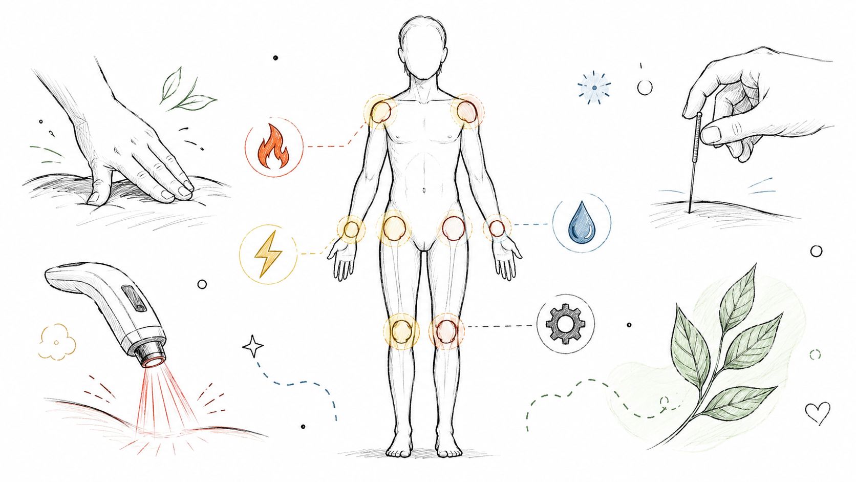
Confused about spinal stenosis vs sciatica? Learn the key differences in symptoms, causes, and treatments to find the right care path in Salt Lake City.
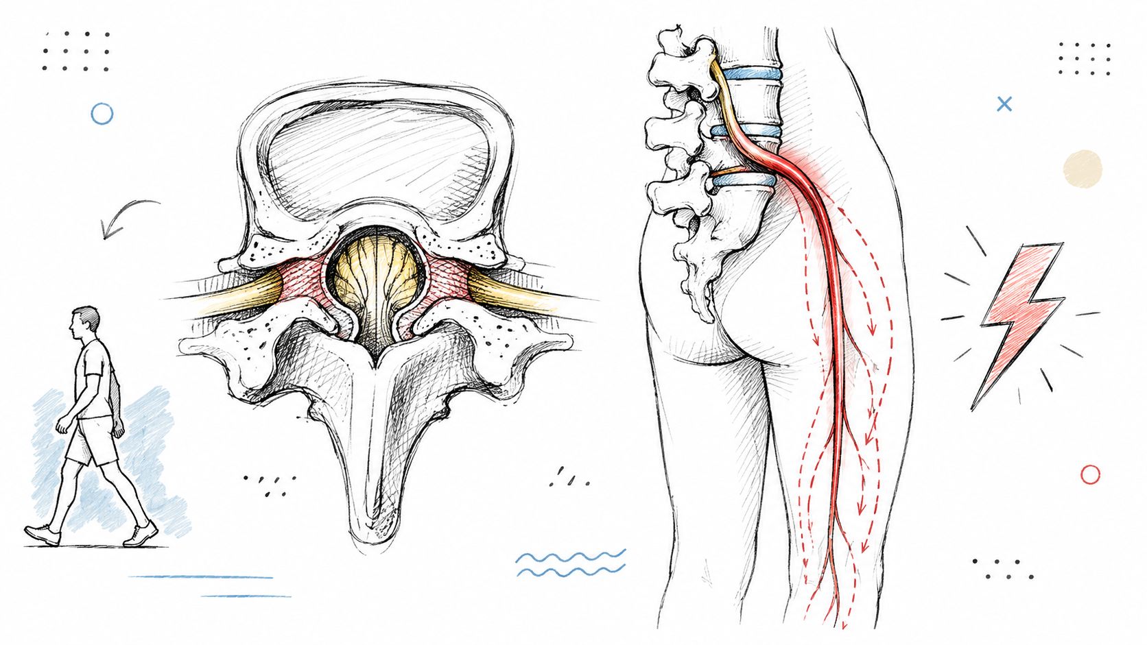



You may be sitting comfortably, yet the moment you stand and walk, pain, tingling, or heaviness starts in your back and travels into your leg. Or perhaps your symptoms feel more like an electric jolt from the buttock down one side, aggravated by sitting, bending, coughing, or sneezing. Patients often ask, “Is this spinal stenosis or sciatica?” The clinically accurate answer is sometimes both.
The difference matters because spinal stenosis is an anatomical diagnosis, while sciatica is a symptom pattern caused by nerve irritation. Identifying whether symptoms come from central canal narrowing, focal nerve-root compression, or overlapping conditions helps determine which examination findings, imaging, and non-surgical treatments make sense.
A patient may arrive with an MRI report describing “stenosis” and say, “So I have sciatica.” Another may describe shooting leg pain and assume the pain itself proves a narrowed spinal canal. Neither conclusion is automatic.
Lumbar spinal stenosis means the space available for the nerves has narrowed. The narrowing may involve the central spinal canal, the side recesses, or the openings where individual nerve roots exit. Age-related changes can contribute to this structural reduction in space, but an image alone doesn't tell us whether the narrowing is causing symptoms.
Sciatica, by contrast, describes a pattern of pain or other nerve symptoms that travels from the lower back or buttock into the leg. It isn't one single disease. Disc herniation is a common cause, but spinal stenosis and other compression mechanisms can also irritate a lumbar nerve root. Historical medical descriptions recognized sciatica as a nerve-related problem, and Domenico Cotugno described its neurogenic origin in 1764, according to this review of sciatica's history and epidemiology.
Practical rule: A scan can show narrowing, but your movement pattern and neurological examination help determine whether that narrowing is clinically important.
The overlap becomes easier to understand with a typical example. Suppose a person has degenerative narrowing around several lumbar nerve roots. Standing upright reduces available space and brings on aching, tingling, or weakness in both legs. That is a stenosis-related pattern, often called neurogenic claudication. If one of those roots becomes especially irritated, the same person may also develop sharper, more localized radiating pain that resembles sciatica.
A disc problem can create the reverse situation. A focal disc herniation may irritate one nerve root and produce classic sciatica without central canal stenosis being the primary issue. A review reports that lumbosacral disc herniation with nerve-root compression accounts for approximately 90% of sciatica cases, with L4-L5 and L5-S1 among the most common levels, as described in this clinical review of sciatica presentation and causes.
Stenosis is common on imaging, especially with age. A systematic review estimated pooled spinal stenosis prevalence at about 11% in the general population and 25% to 39% in clinical settings, while radiologic narrowing reached about 38% in the general population. The same review noted radiologic stenosis in about 11% of asymptomatic people, showing why a scan must be interpreted alongside symptoms and examination findings. See the systematic review of lumbar spinal stenosis prevalence.
That doesn't mean imaging is unhelpful. It means the useful question isn't just, “Do I have narrowing?” It's, “Does the location and severity of the narrowing match what happens when I stand, walk, sit, bend, or move?”
The most useful distinction in daily life is often what triggers the symptoms and what relieves them. Stenosis-related neurogenic claudication usually depends on standing, walking, and lumbar extension. Sciatica from focal nerve-root irritation more often follows a sharp, shooting path into one leg and may be influenced by sitting, bending, coughing, or sneezing.
The patterns can overlap, so the table below is a guide, not a diagnosis.
| Feature | Spinal Stenosis | Sciatica |
|---|---|---|
| Primary description | Anatomical narrowing around spinal nerves | Symptom pattern caused by nerve irritation |
| Typical trigger | Standing or walking | Sitting, bending, coughing, sneezing, or certain spinal movements may aggravate symptoms |
| Common relief | Sitting or leaning forward | Varies according to the irritated nerve root and underlying cause |
| Pain quality | Aching, burning, cramping, heaviness, or leg discomfort | Sharp, shooting, electric, or radiating pain |
| Distribution | One or both legs, sometimes with one side worse | Often follows a more specific pathway in one leg |
| Neurological findings | May be normal or equivocal unless a focal root is also compressed | Numbness, weakness, or reduced reflexes may occur |
| Walking tolerance | Often reduced because symptoms build with activity | Can be limited by pain, weakness, or nerve sensitivity |
A person with stenosis may say, “I can walk through a grocery store only if I lean over the cart.” That forward-flexed position commonly feels better because lumbar flexion can reduce the activity-related irritation associated with narrowed spaces. Standing tall or arching backward tends to be more provocative.
The symptoms may include buttock or leg pain, paresthesia, cramping, numbness, and weakness. The clinical overview of lumbar spinal stenosis symptoms identifies burning or aching pain into the buttocks and legs, along with numbness, tingling, cramping, and weakness, as common features.
Sciatica often feels more sharply defined. A patient may point from the lower back into the buttock and down the thigh or calf, describing a narrow, shooting route. If a particular root is involved, the examination may reveal sensory loss in a corresponding area, weakness in a related muscle group, or a reduced reflex.
Pain distribution alone still isn't enough. Some people with stenosis have one-sided symptoms, and some people with sciatica have symptoms that don't follow a perfect textbook line. During evaluation, describe the exact activity that starts the pain, how long it takes to appear, whether sitting helps, and whether coughing or sneezing changes it.
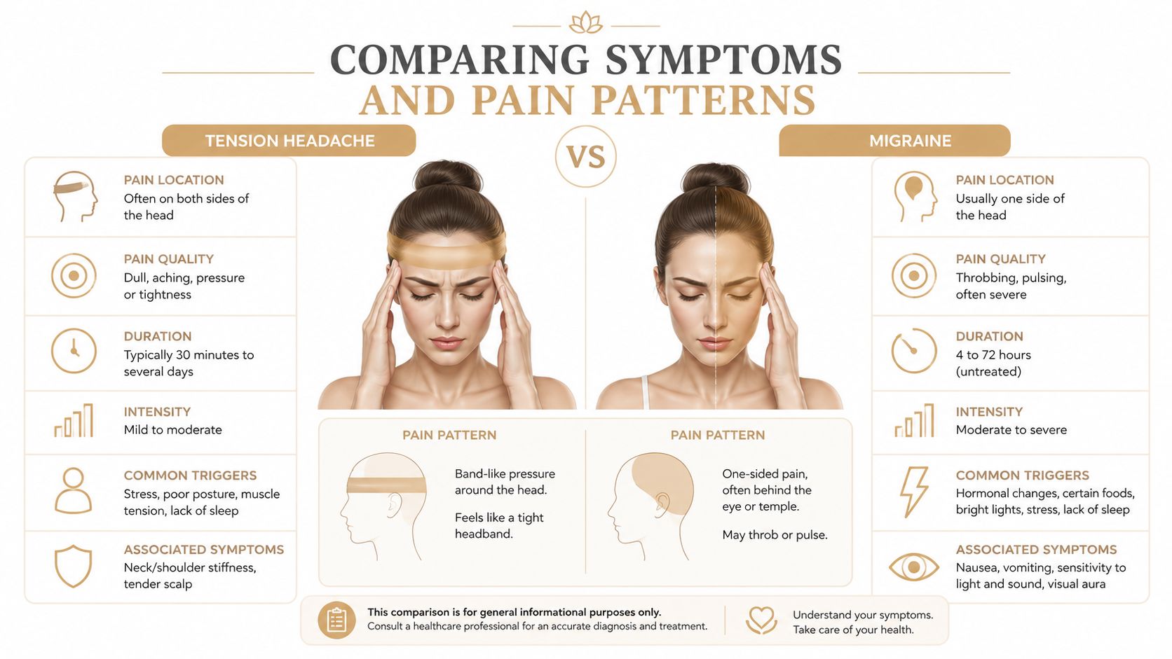
People who run or stay active often need a separate discussion about load, stride, hip mobility, and trunk control. A practical resource on strength drills for back pain runners can help frame that conversation, but persistent radiating pain, weakness, or walking limitation deserves an individual assessment rather than a generic exercise list.
A reliable diagnosis begins with the history, then tests whether the symptoms match the examination. Ask whether pain or heaviness appears with walking, standing, sitting, bending, or coughing. Clarify whether one or both legs are involved and whether function is declining. Examination should compare strength, sensation, reflexes, gait, spinal and hip movement, and nerve-tension responses.
Lumbar stenosis can produce a relatively normal or equivocal neurological examination, particularly when narrowing affects several structures without severe focal nerve-root dysfunction. Neural-tension tests may also remain negative unless one root is specifically compressed.
Sciatica or lumbosacral radicular pain more often produces objective findings such as hypoesthesia, paresis, and diminished reflexes, although a patient may not show all three. A sound assessment combines the history and examination with targeted investigation rather than treating pain location as proof of one diagnosis. The evidence is discussed in this review of diagnostic assessment for lumbar radicular pain.
The clinical overlap matters. A narrowed canal can irritate multiple nerve structures, while a disc or foraminal problem can create a more focal nerve-root pattern. Imaging may show both processes, so the treatment plan should address the mechanical narrowing and the irritated nerve rather than force every symptom into a single category.
X-rays can show alignment, degenerative changes, vertebral height, and other bony features. They provide limited information about nerve roots and soft tissues. MRI is more useful when symptoms persist, are severe or progressive, or include neurological deficits because it can show discs, the central canal, lateral recesses, and foraminal spaces.
Some payer policies require about 6 weeks of conservative care before MRI for suspected stenosis when red flags are absent. That timing supports a function-based evaluation instead of automatic scanning after every episode. The clinical reference on lumbar spinal stenosis emphasizes matching imaging findings to the clinical picture. For a deeper comparison of scan selection, see this guide on MRI vs X-ray for back pain.
One clinical comparison found spinal canal stenosis in 37% of patients with sciatica and 11% of patients with low back pain. Imaging signs of nerve-root compression were also more common in the sciatic group, including 91% with at least one finding of compression. These figures come from the study comparing low back pain and sciatica imaging findings. They support careful interpretation. An abnormal scan does not automatically identify the pain generator.
Seek urgent medical evaluation for new bowel or bladder dysfunction, saddle-area numbness, rapidly worsening weakness, major loss of coordination, fever with severe back pain, or serious trauma. These findings may indicate a condition requiring medical assessment beyond routine conservative care.
Treatment should match the driver of symptoms, the examination, irritability, and functional goals. A plan for focal sciatica may emphasize reducing nerve sensitivity and improving tolerance to movement. A plan for stenosis may focus more on flexion-tolerant positions, walking capacity, hip and trunk control, and strategies that reduce repeated extension loading. When both patterns appear, combining approaches is often more sensible than forcing the patient into one diagnostic category.
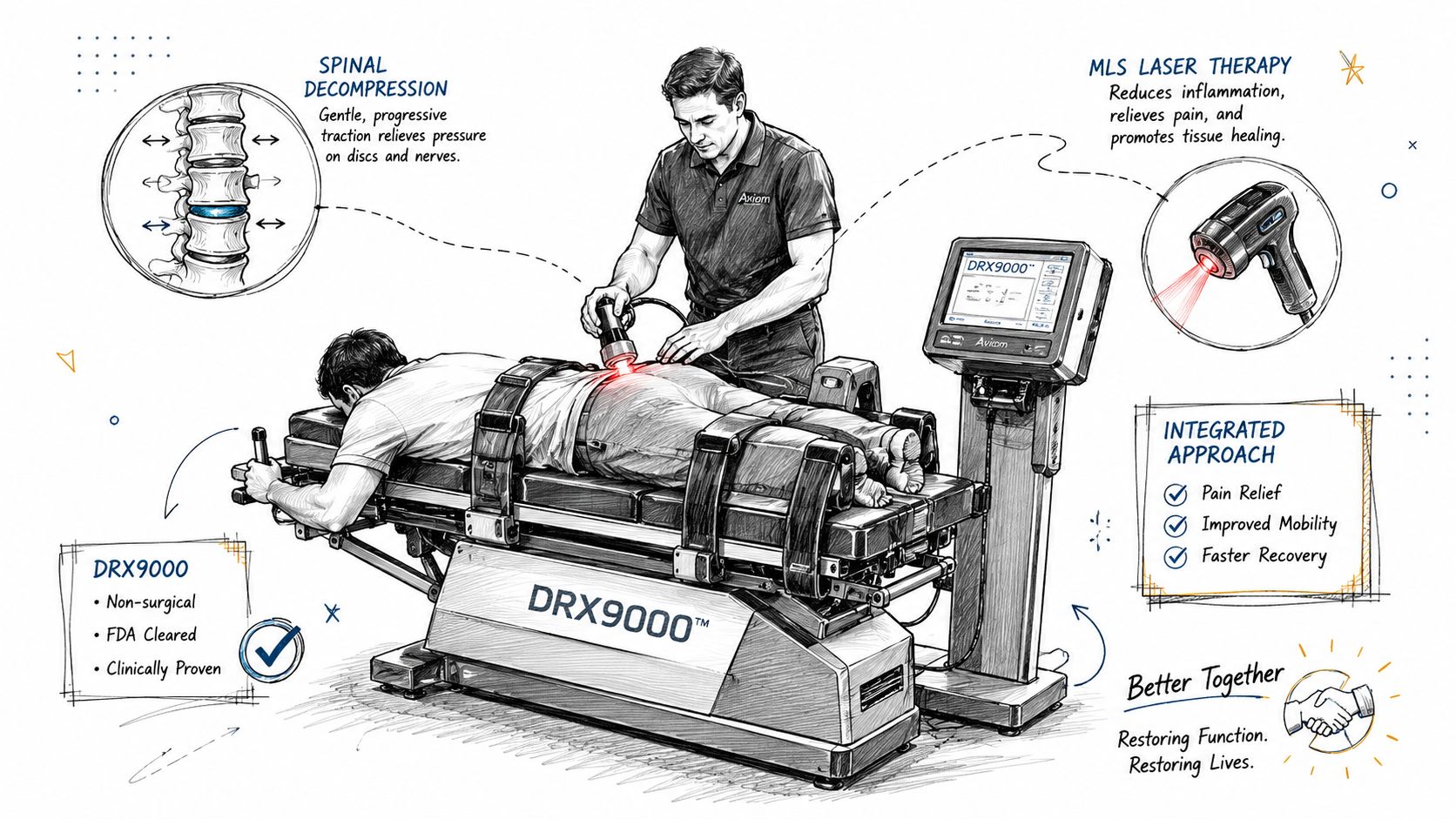
At Aspen Falls Wellness, care may include chiropractic adjustment, spinal decompression, rehabilitation exercise, and supportive modalities within an individualized plan. The purpose isn't to apply every service to every patient. The purpose is to select the tools that address the mechanical, neurological, muscular, and functional pieces of the presentation.
Before choosing treatment, establish what the patient can't do. Is the limiting problem standing long enough to cook, walking across a parking lot, sleeping, lifting, or returning to golf? A baseline makes it easier to judge whether care is improving function rather than only producing temporary table relief.
A practical plan can include:
The overview of spinal decompression therapy explains how decompression can fit into a non-surgical care discussion.
Passive treatment can make movement easier, but lasting progress depends on what the patient can do between visits. Mobility Therapy and posture retraining can improve movement options, while Rehabilitation Exercise builds tolerance for walking, lifting, and daily activity. Acupuncture may help with pain modulation, and Nutrition and Nutrition Counseling can support broader recovery habits without pretending that diet alone can enlarge a narrowed spinal canal.
For active adults, a Golf Movement Screening can identify swing or rotation demands that repeatedly irritate the back. For people recovering after a collision, Car Accident Treatment begins with a careful assessment because trauma changes the clinical context. The common thread is re-evaluation. If leg weakness, walking tolerance, or neurological signs worsen, the plan should change and referral may be appropriate.
The long-term outlook is more nuanced than “stenosis always gets worse” or “sciatica always disappears.” Symptoms can improve, fluctuate, or plateau, and the structural findings on imaging don't move in perfect step with pain. The right target is a safer, more capable daily life, supported by follow-up when the pattern changes.
A 2026 long-term study found low-certainty evidence that people with lumbar spinal stenosis treated non-surgically may improve by up to about one-third in pain and disability during the first year, with little additional improvement through five years. The results are reported in this long-term study of non-surgical lumbar spinal stenosis outcomes. That doesn't predict one person's outcome, but it does challenge the expectation that conservative care will produce uninterrupted improvement indefinitely.
Pain scores matter, but they shouldn't be the only marker. A patient who still has some discomfort but can walk farther, sleep better, stand to prepare a meal, or return to controlled recreational activity may be making meaningful progress.
Useful goals include:
Sciatica from disc herniation often has a favorable natural course, and one PubMed review reports favorable spontaneous outcomes in 95% of cases caused by disc herniation. The same evidence base also notes that severe pain can occur without clear imaging compression and that pain severity doesn't reliably match herniation size, as described in this review of sciatica prognosis and treatment. These findings support conservative care for many patients, while progressive weakness or other warning signs still require timely medical attention.
Once acute irritability settles, mobility work, strengthening, walking strategy, posture training, and nutrition habits can help maintain capacity. This isn't a promise that maintenance care eliminates degeneration. It gives the patient more control over the factors that influence movement tolerance and recurrence.
Progressive rehabilitation should be adjusted when symptoms plateau. A plateau isn't a reason to keep repeating the same passive treatment indefinitely. It may be time to reassess the diagnosis, modify exercises, review imaging, or seek a specialist opinion.
Consider a professional evaluation when leg symptoms limit walking, standing, work, sleep, exercise, or household activity. You should also seek assessment when symptoms persist despite basic conservative measures, return repeatedly, or include numbness, weakness, altered reflexes, or a noticeable decline in function.
A coordinated team can be useful when the presentation includes both stenosis-related activity intolerance and focal sciatica-like nerve symptoms. Chiropractic Care can address joint motion and mechanical contributors. Acupuncture and Massage Therapy may support pain modulation and soft-tissue comfort. Mobility Therapy and Rehabilitation Exercise can translate symptom relief into improved walking, lifting, and recreational capacity.
A useful consultation should answer practical questions:
Residents of Salt Lake City and Sandy can seek this type of multidisciplinary, non-surgical evaluation through a local practice that coordinates chiropractic, acupuncture, massage, mobility, nutrition, and rehabilitation services. The guide to chiropractic care for sciatic nerve pain can help you understand what a conservative evaluation may involve, but an in-person examination remains important when symptoms are persistent or complex.
Don't wait for leg pain to define your routine. Visit Aspen Falls Wellness for an individualized evaluation addressing spinal stenosis, sciatica, mobility, and function, with options including Chiropractic Adjustment, Spinal Decompression with the DRX 9000, MLS Laser Therapy, SoftWave Therapy, acupuncture, massage, and rehabilitation exercise. Bring a clear description of what triggers and relieves your symptoms so the team can build a focused, non-surgical care plan.

Stay updated with the latest chiropractic news and exclusive offers.