Learn how is spinal stenosis diagnosed through history, exam maneuvers, imaging, and red flags. A practical guide for patients and clinicians.
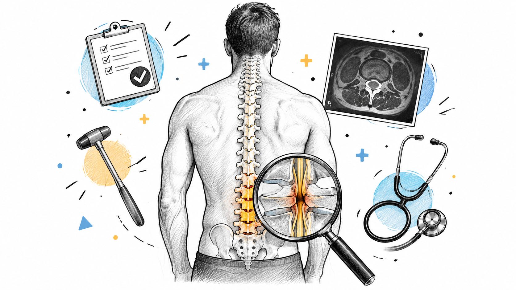
The most popular advice about spinal stenosis is also the most misleading: “Get an MRI.” An MRI can show narrowing, but it can't decide whether that narrowing explains your pain, limits your walking, or requires treatment. Spinal stenosis is diagnosed by matching a clinical pattern to physical findings and then using imaging to confirm the anatomy.
In a Salt Lake City chiropractic practice, the most useful clues often appear before a scan is ordered. I want to know what happens when you stand, how far you can walk, whether sitting changes your symptoms, and whether your strength, reflexes, sensation, and gait have changed. That history determines whether imaging is useful, whether another condition is more likely, or whether you need prompt referral.
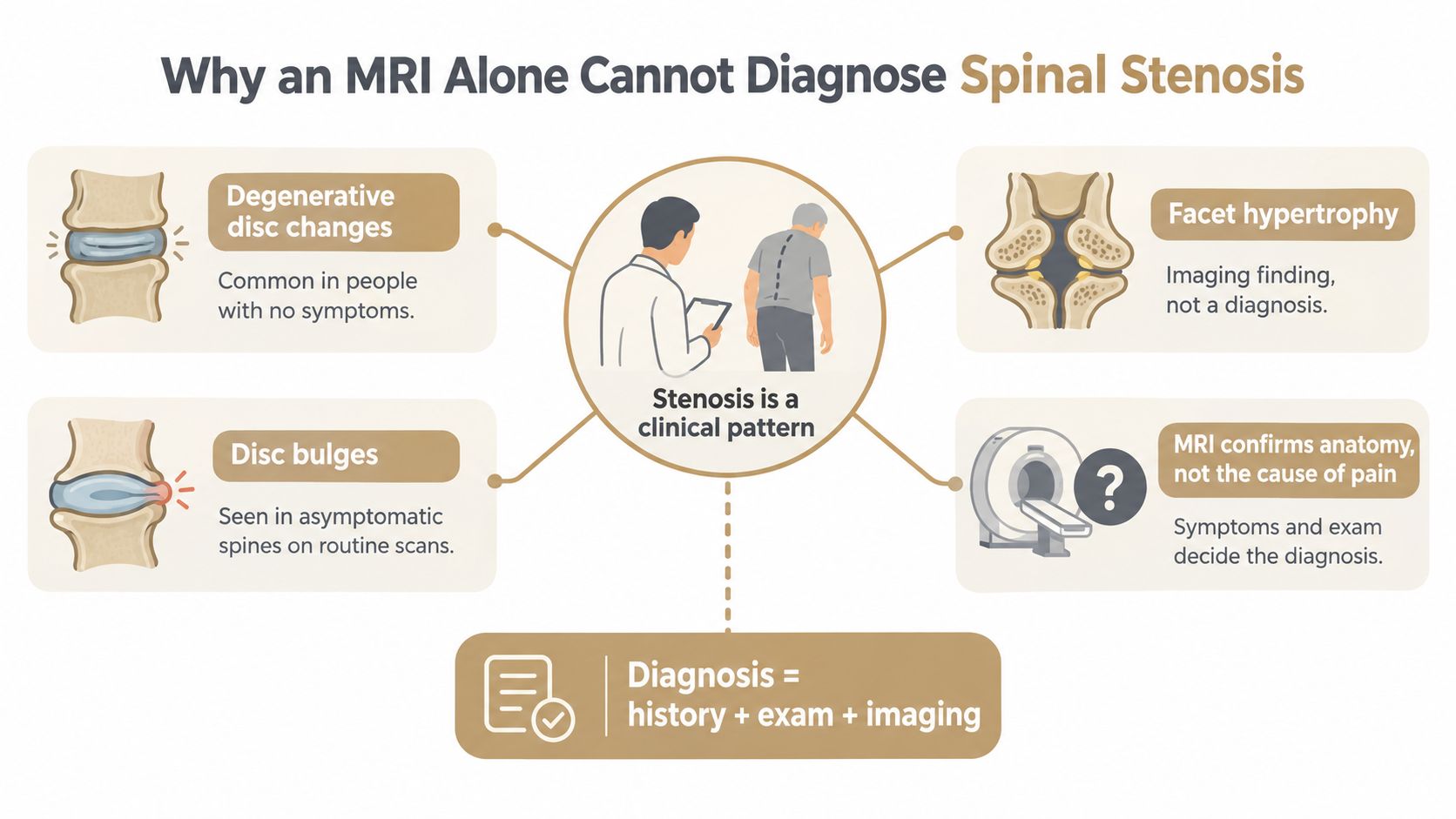
An MRI alone cannot diagnose clinically meaningful spinal stenosis. It can identify a narrowed canal, crowded nerve roots, disc changes, thickened ligaments, or enlarged facet joints. Those findings describe anatomy, but they don't automatically identify the source of pain.
Radiologic narrowing is common in people who don't have symptoms. One review found central canal stenosis in 14.0% of asymptomatic people and 59.5% of symptomatic people, while another prevalence study reported lumbar stenosis in 11% of asymptomatic people and 38% of the general population when defined by imaging findings (review of imaging and clinical correlation). The practical lesson is simple: an abnormal MRI can be relevant, incidental, or only one part of a larger problem.
The strongest clinical pattern is neurogenic claudication. Leg pain, heaviness, numbness, or weakness tends to worsen with standing or walking and improve with sitting or forward flexion. A patient who can tolerate leaning over a shopping cart but struggles to stand upright is giving the clinician useful mechanical information.
That history matters because imaging severity correlates only weakly with symptom intensity. Two people can have similar narrowing on MRI and very different levels of disability. A person with a dramatic-sounding report may walk comfortably, while someone with less striking imaging may be unable to stand long enough to cook or shop.
Practical rule: Treat the patient's walking tolerance and neurological findings as clinical evidence. Treat the MRI as an anatomical map.
X-rays and MRI also answer different questions, as explained in this guide to MRI versus X-ray for back pain. The correct sequence is usually history first, examination second, and imaging when the result can confirm or change the working diagnosis. That approach reduces unnecessary worry and makes it less likely that a scan will become the diagnosis by itself.
A careful history often narrows the diagnosis before anyone orders an advanced test. The clinician needs more than the location of your back pain. The timing, triggers, posture, walking tolerance, and associated neurological symptoms reveal how the nerves may be responding to a narrowed space.
The central question is whether your symptoms behave like neurogenic claudication. Tell the clinician whether leg heaviness, cramping, burning, tingling, or weakness appears during standing or walking, whether it improves when you sit, and whether bending forward changes the distance you can travel.
Standing and walking aren't interchangeable clues. Some people notice symptoms from standing upright, while others can stand briefly but develop leg symptoms after walking. Relief from sitting or lumbar flexion supports a spinal pattern. Leg pain that continues regardless of posture raises concern for another cause, including a circulation-related problem.
The timeline matters, too. A gradual change in walking tolerance suggests a different workup from sudden symptoms after trauma. Mention prior back surgery, injuries, work demands, positions that aggravate symptoms, and any change in balance. Report numbness in the groin region, bowel or bladder changes, or rapidly increasing weakness immediately.
| History Question | What a Typical Answer Looks Like | What It Suggests |
|---|---|---|
| What brings on the leg symptoms? | Standing upright or walking makes the legs heavy, painful, numb, or weak | A pattern compatible with neurogenic claudication |
| What relieves the symptoms? | Sitting or bending forward provides relief | Symptoms may be posture-dependent and related to lumbar narrowing |
| Is walking or standing more provocative? | Walking limits distance, while standing may also reproduce symptoms | Helps distinguish the activity and posture that provoke nerve irritation |
| Where do you feel the symptoms? | Both legs, one leg, or a narrower path into the foot | Helps direct attention toward central, lateral recess, or foraminal involvement |
| Did the symptoms start after an event? | A gradual change, injury, surgery, or sudden neurological change | Determines whether degenerative narrowing or another process needs consideration |
| Have bowel, bladder, or saddle symptoms changed? | New retention, incontinence, or numbness around the groin | A warning sign requiring urgent medical assessment |
Patients often describe the “shopping cart” posture without knowing it has diagnostic value. Leaning forward changes the lumbar position, so the response to that posture helps the clinician decide whether the symptoms fit a stenotic pattern. It isn't proof by itself, but it is more useful than asking only how severe the pain feels.
Bring a short symptom record to an appointment. Note what happens during a walk, how long you can stand, whether sitting changes the symptoms, and whether the problem affects one or both legs. Those observations can make the examination and any later imaging more focused.
The examination tests whether the story has a physical correlate. I watch how a person enters the room, rises from a chair, turns, and walks before focusing on isolated muscle tests. A stooped or wide-based gait can raise suspicion, but it isn't specific enough to establish stenosis on its own.
Gait observation may reveal a forward-leaning posture, cautious steps, or balance changes. A wide-based gait can support concern about neurological involvement, while a normal Romberg test doesn't rule out lumbar stenosis. Single-leg stance challenges balance and lower-limb control, but inability to hold the position can also reflect hip, vestibular, or general strength problems.
Repeated toe-walking and heel-walking provide a quick screen of lower-leg strength. Weakness with heel-walking can point toward reduced dorsiflexion strength, while difficulty with toe-walking may indicate a different lower-leg motor deficit. These findings help identify a nerve-related problem, but they don't locate every source of symptoms by themselves.
A straight-leg raise is useful when it reproduces familiar radiating leg pain, particularly when a disc-related nerve irritation is being considered. Its value is more limited in central spinal stenosis, where walking-related heaviness and posture dependence may be more informative than a resting leg-raise maneuver.
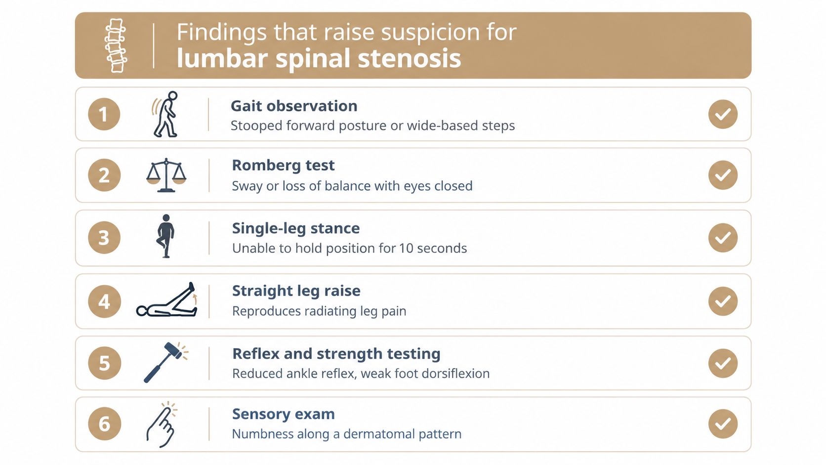
A focused neurological examination compares sensation across the lower-limb dermatomes, checks patellar and Achilles reflexes, and tests key muscle groups. A sensory change that follows a nerve-root pattern carries more weight than vague, symmetrical foot discomfort. Reflex asymmetry or weakness can support nerve involvement and help determine whether referral or imaging should be accelerated.
A clinician may also use a short treadmill or walking provocation assessment. Reproduction of familiar leg symptoms with lumbar extension, followed by relief while sitting or leaning forward, strengthens the case for neurogenic claudication. The test is not a standalone diagnosis, and it should stop if symptoms become unsafe.
Lower-extremity pulse checks are important when leg pain occurs with walking. Reduced pulses or symptoms that don't vary with posture can shift attention toward vascular causes. A first chiropractic visit should include a clear explanation of what is being tested and why, not just a series of movements without context.
Clinical observation: A normal strength test at rest doesn't guarantee normal walking function. Some patients develop their most important symptoms only after standing or walking.
Physical examination and X-rays have limited sensitivity and specificity when used alone. Cross-sectional imaging is commonly needed when the history and examination point toward clinically relevant stenosis, as summarized in this clinical review of lumbar spinal stenosis diagnosis.
Imaging is most useful when the history and examination suggest that the result could change care. It isn't automatically necessary for every episode of back pain, and it shouldn't replace a neurological examination.
MRI is usually the preferred advanced test for suspected lumbar spinal stenosis. It shows the spinal canal, nerve roots, discs, ligaments, and other soft tissues. It can help identify central canal, lateral recess, or foraminal narrowing and distinguish degenerative narrowing from conditions such as tumor, infection, hematoma, disc disease, or ligament disease (clinical overview of spinal stenosis diagnosis).
Plain radiographs are useful for assessing alignment, bone changes, disc-space height, spondylolisthesis, and previous hardware. They don't show the spinal cord, nerve roots, or soft-tissue compression well, so a normal or nonspecific X-ray doesn't exclude stenosis.
MRI provides the most complete noninvasive view of the structures that may crowd the canal or nerve exits. It should be ordered when the clinical pattern is convincing and the result may influence rehabilitation, injections, surgical consultation, or another major decision. Guidance on lumbar stenosis identifies MRI as the appropriate test when confirmation of anatomic narrowing or nerve-root impingement is needed (lumbar spinal stenosis imaging guidance).
CT or CT myelography can provide additional detail about bone, the canal, and nerve-root pathways. CT myelography may be considered when MRI isn't possible or when more precise anatomical detail is needed for decision-making. The trade-off is that CT-based studies are less informative than MRI for many soft-tissue structures and may involve additional procedural considerations.
| Modality | Best For | Limitations |
|---|---|---|
| X-ray | Alignment, bone changes, disc height, instability clues, and hardware review | Doesn't show nerve roots or soft-tissue compression adequately |
| MRI | Canal narrowing, discs, ligaments, nerve roots, and other soft tissues | Findings may be present without symptoms and must match the examination |
| CT | Detailed bony anatomy and complex structural review | Less effective for soft-tissue detail than MRI |
| CT myelography | Canal and nerve-root detail when MRI isn't possible or more detail is required | Invasive compared with routine MRI and not the first choice for every patient |
A practical imaging threshold sometimes used for lumbar stenosis is a canal measurement of 12 mm or less, while a threshold under 10 mm is often cited for cervical stenosis (radiology guidance on imaging spinal stenosis). These measurements can describe narrowing, but they don't determine symptom severity or automatically indicate surgery. The diagnosis still requires both radiologic evidence and compatible clinical symptoms (review of diagnostic criteria and treatment implications).
Leg pain with walking doesn't belong to the spine by default. The clinician compares posture, walking behavior, pulse findings, sensory distribution, joint motion, and neurological results before deciding that stenosis is the best explanation.
Vascular claudication may also cause cramping or aching during activity. The key distinction is often what happens when the person stops. Neurogenic symptoms tend to improve with sitting or forward flexion, while vascular symptoms may improve just by standing still. Pulse assessment and appropriate vascular testing can help separate the two patterns.
Peripheral neuropathy usually produces a more distal and symmetrical pattern of numbness or burning that doesn't change reliably with lumbar posture. A clinician may consider laboratory or electrodiagnostic evaluation when the history and examination suggest a peripheral nerve process rather than compression in the spinal canal.
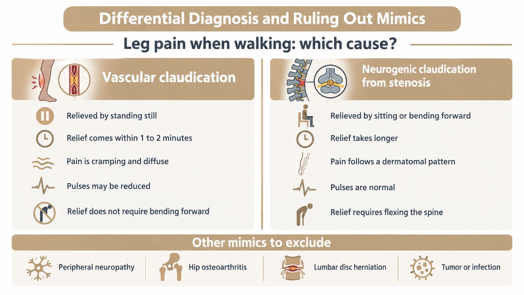
Hip osteoarthritis can refer pain into the thigh or buttock and often limits hip movement, particularly internal rotation. A disc herniation may produce more acute, sharp, radiating pain and a positive straight-leg raise, without the characteristic walking-and-posture pattern of central stenosis.
Other neurological or systemic conditions can mimic stenosis, including diabetic nerve disorders, inflammatory disease, spinal tumors, and infection. The workup may therefore include targeted imaging, blood tests, or nerve studies when the presentation doesn't fit a straightforward degenerative pattern.
| Pattern | More consistent with | What needs checking |
|---|---|---|
| Leg symptoms improve with sitting or forward flexion | Neurogenic claudication | Lumbar examination and, when indicated, MRI |
| Symptoms improve by stopping activity without needing to bend forward | Vascular claudication | Pulses and vascular assessment |
| Symmetrical distal burning or numbness with little posture variation | Peripheral neuropathy | Medical history, laboratory assessment, or nerve testing |
| Groin pain with restricted hip movement | Hip disorder | Hip examination and targeted imaging |
| Sharp, acute radiating pain with a positive straight-leg raise | Disc-related nerve irritation | Neurological examination and appropriate imaging |
A diagnosis of exclusion is still a diagnosis, but only after serious and treatable alternatives have been considered. This is why spinal stenosis versus sciatica shouldn't be treated as a simple label choice. The same patient can have overlapping findings, and the treatment plan depends on which pattern is limiting function.
Most suspected degenerative stenosis can be evaluated in a planned manner. Certain symptoms change that pace immediately because they may indicate serious neurological compromise rather than routine narrowing.
New bowel or bladder retention, loss of control, or numbness around the saddle region requires urgent medical assessment. Progressive weakness in both legs, rapidly worsening motor loss, or marked gait deterioration also warrants prompt imaging and specialist evaluation. These findings can occur with compression of the cauda equina or spinal cord and shouldn't wait for a routine appointment.
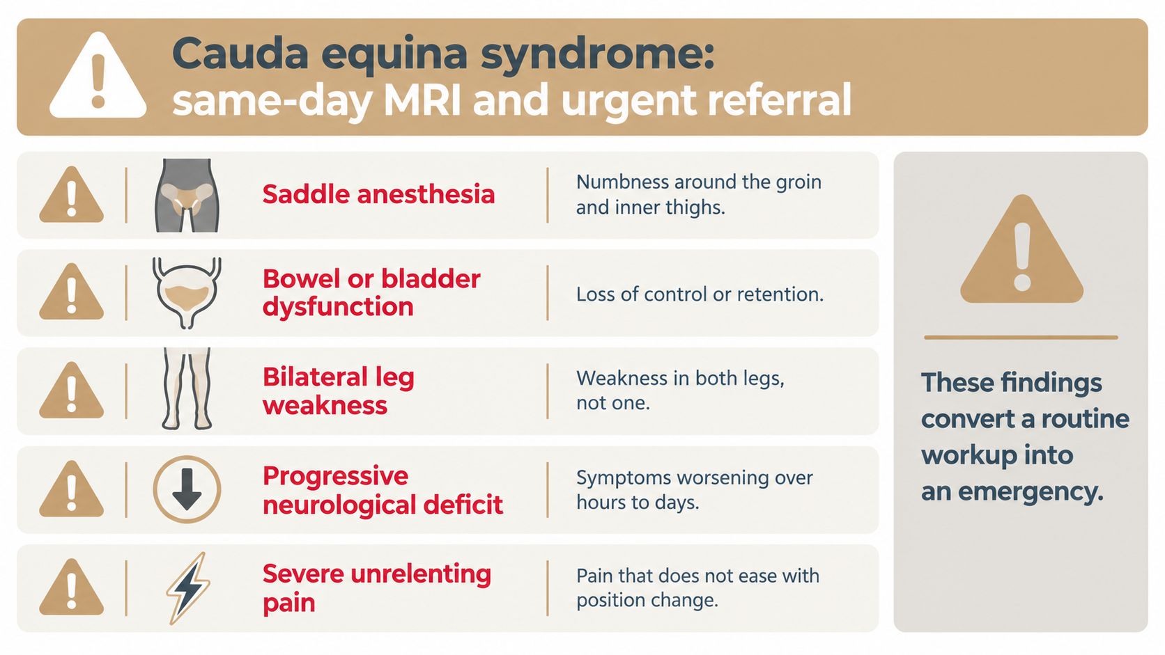
Seek urgent care for:
Recent severe trauma can raise concern for fracture. Fever, a history of cancer, immune suppression, or other systemic warning signs may point toward infection or malignancy rather than ordinary degenerative stenosis. The clinician may then arrange accelerated MRI, laboratory evaluation, and specialist referral.
Urgent distinction: Saddle numbness or bowel and bladder dysfunction isn't a routine chiropractic adjustment problem. Go to an emergency setting rather than waiting for conservative treatment to clarify the symptoms.
A spinal stenosis algorithm from the McGill Spine Program identifies bowel or bladder dysfunction, major trauma, progressive weakness, and other neurological signs as reasons for faster investigation. A chiropractor can recognize these warning signs, but the appropriate next step is referral for medical assessment.
A diagnosis should answer two questions: what structure is involved, and what function has been lost? Treatment should then target walking tolerance, standing capacity, sleep, balance, and daily activity rather than trying to make an MRI report look different.
For patients without urgent neurological findings, conservative care commonly begins with activity modification and a structured rehabilitation plan. Flexion-tolerant movement, trunk and hip strengthening, mobility work, posture retraining, and carefully progressed walking can help patients stay active without repeatedly provoking symptoms. Manual therapy may be appropriate when joint or soft-tissue restrictions are contributing, but it should be adapted to the person's neurological findings and tolerance.
Medication decisions belong with a qualified medical professional. In selected cases, an epidural injection may provide a time-limited bridge that makes rehabilitation more manageable. It doesn't correct the underlying anatomy, so its value should be judged by whether it improves function and supports a broader plan.
Progressive neurological deficits, emergency red flags, or persistent functional limitation despite a well-delivered conservative plan warrant a spine specialist consultation. Imaging alone doesn't require surgery, and a surgical discussion doesn't obligate anyone to have an operation. The purpose of referral is to clarify options when symptoms, examination, and anatomy point in the same direction.
At Aspen Falls Wellness, care may include chiropractic adjustment, mobility therapy, rehabilitation exercise, massage therapy, acupuncture, SoftWave Therapy, MLS Laser Therapy, nutrition counseling, and personalized home exercise plans. Spinal Decompression with the DRX 9000 may be considered for selected disc-related presentations, but it isn't a substitute for urgent referral when neurological red flags are present.
The same principle applies to realignment, muscle stimulation, pain relief programs, and sciatica treatment. These services should follow an examination and a defined functional goal. Re-imaging usually isn't the next move when symptoms are stable and the plan is working. New neurological findings or a meaningful change in symptoms can justify another evaluation.
Diagnosis is the start of a plan, not the end of the workup. If your walking distance is shrinking, your legs feel unreliable, or sitting is the only reliable relief, arrange an evaluation that connects your history, examination, and imaging instead of relying on an MRI label.
If back or leg symptoms are limiting your walking, Aspen Falls Wellness can evaluate your movement and neurological findings, recommend imaging referral when indicated, and build a non-surgical care plan around your goals. Contact the Salt Lake City or Sandy clinic to discuss whether chiropractic care, mobility therapy, rehabilitation exercise, or another appropriate service fits your presentation.

Stay updated with the latest chiropractic news and exclusive offers.