Explore proven disc herniation treatment without surgery options, timelines, red flags, and how integrative care can relieve pain and restore function.
Many people assume a lumbar disc herniation is a straight line to surgery. The long-term record says otherwise. In one classic follow-up of 30 patients with massive prolapsed discs treated conservatively, 17 had complete resolution, 10 had only occasional symptoms, and overall satisfaction reached 90% in the non-surgical group, according to the Royal College of Surgeons review. That's the right starting frame for disc herniation treatment without surgery: not denial, not wishful thinking, but a structured trial with close monitoring and a clear threshold for escalation.
Most public advice stops at “rest and try therapy.” That's too vague to be useful. The job is separating pain-only cases from cases with progressive neurologic loss, then building a plan that reduces nerve irritation, maintains function, and checks whether the leg is recovering.
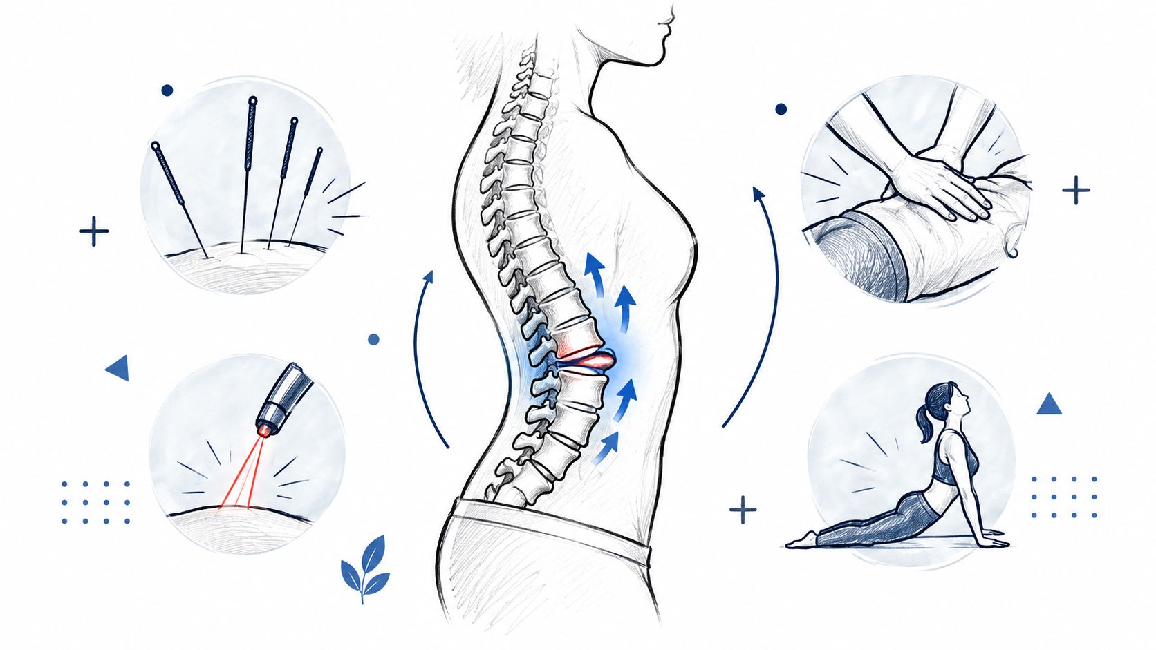
A lumbar disc herniation happens when the annulus fibrosus, the outer ring of the disc, weakens or tears and the nucleus pulposus, the softer inner material, pushes outward. If that displaced material irritates or compresses a nearby nerve root, people usually feel more than back pain. They feel radiating leg pain, numbness, tingling, or weakness that makes sitting, driving, bending, and sleep miserable.
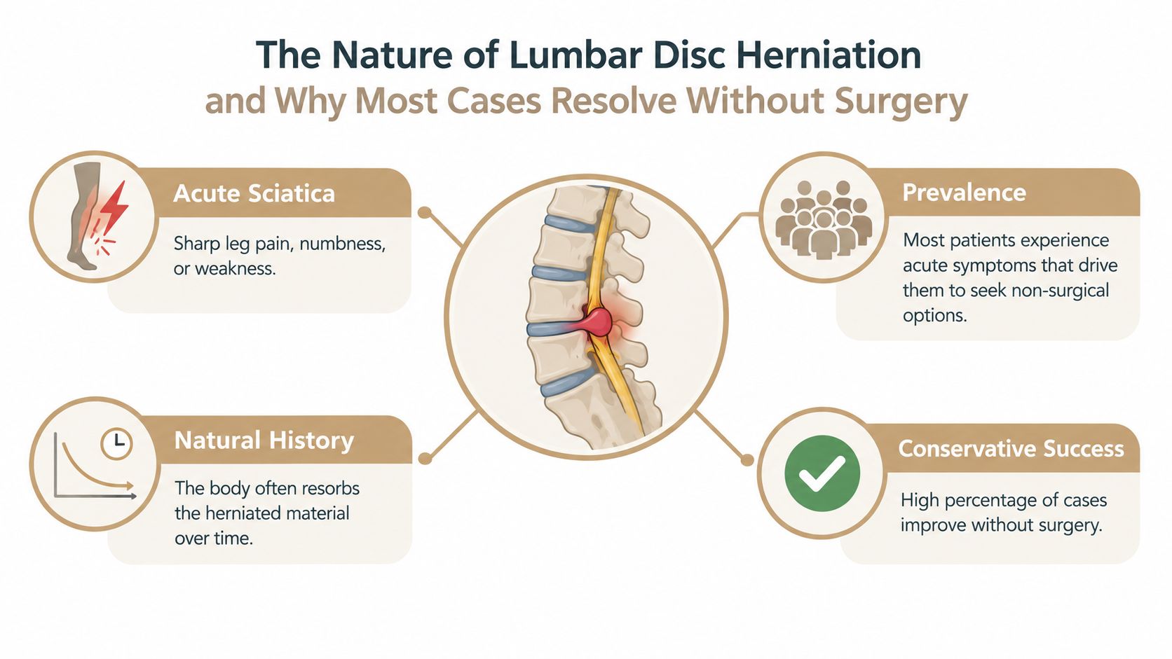
Patients often get stuck on the wording in the scan report. The terms matter, but they aren't a verdict.
Those labels help a clinician match the image to the exam. They don't automatically decide treatment.
When there's no cauda equina syndrome and no progressive motor deficit, first-line care is conservative. Major clinical summaries recommend a practical sequence of red-flag screening, light activity instead of prolonged bed rest, symptom control with NSAIDs or analgesics as needed, then physical therapy and home exercise. They also note that many symptomatic cases improve within 6 to 8 weeks, and a conservative trial of about 4 to 12 weeks is commonly recommended before surgery is considered if pain remains intractable or neurologic deficits persist, as summarized in this clinical review on lumbar disc herniation management.
Clinical reality: The body often settles the inflammatory storm before anyone needs an operation. The mistake is either panicking too early or waiting too long when weakness is getting worse.
This is why disc herniation treatment without surgery works best when it's structured. The disc can calm down, the fragment may become less irritating over time, and the nerve can recover. But that only helps if someone is tracking function, not just pain.
A good disc workup starts with the history, not the image. The first questions are practical. Where does the pain travel? Does it go below the knee? Is there numbness in the shin, top of the foot, outer calf, or sole? Did the problem start after lifting, twisting, coughing, or did it appear without a clear trigger? Are bowel or bladder changes present? Is pain waking the patient at night?
A focused lumbar and neurologic exam can identify the involved level in many cases before any imaging is ordered. The essentials include gait, straight-leg raise, slump testing, femoral stretch testing when upper lumbar involvement is suspected, myotomal strength, dermatomal sensation, and reflexes.
| Clinical Exam Findings by Disc Level | Pain Radiation | Weakness | Sensation Loss | Reflex Change |
|---|---|---|---|---|
| L3-L4 | Front of thigh, sometimes toward knee | Quadriceps weakness may appear | Anterior thigh or medial knee region | Patellar reflex may change |
| L4-L5 | Lateral leg, top of foot | Ankle dorsiflexion or big toe extension may weaken | Dorsum of foot, lateral shin | Reflex findings may be subtle |
| L5-S1 | Back of leg, outer foot, sole | Plantarflexion may weaken | Lateral foot or sole | Achilles reflex may change |
If you want a symptom-by-symptom breakdown of how these patterns show up, this guide to lumbar disc herniation signs and symptoms is a useful companion.
MRI is most useful when red flags are present, when symptoms persist despite a structured trial of care, when weakness is progressing, or when surgical planning is on the table. Plain X-ray can help if fracture, alignment change, or degenerative instability is part of the question. CT has a narrower role, usually when MRI isn't available or isn't appropriate.
Many people have disc findings on imaging that don't match their symptoms. The exam decides whether the image matters.
That point saves people from overtreatment. Imaging should confirm a working diagnosis, not replace one.
No single therapy carries uncomplicated lumbar disc cases by itself. The best conservative plans usually combine mechanical unloading, pain modulation, movement restoration, and home exercise. What matters most is using each tool for the right job and at the right stage.
| Non-Surgical Disc Herniation Therapies Compared | Primary Target | Typical Cadence | Best-Fit Patient |
|---|---|---|---|
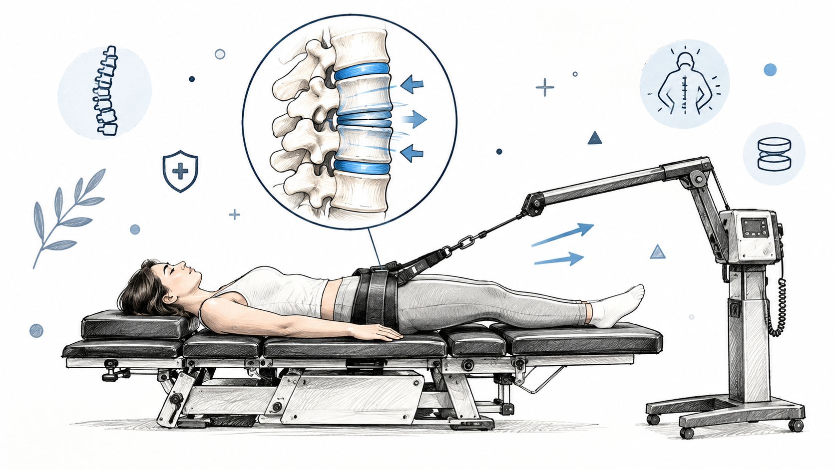
| Spinal Decompression | Mechanical unloading and reduced nerve irritation | Often used several times weekly early in care | Patients with disc-related leg pain who are aggravated by compression and sitting |
| Chiropractic Adjustment | Segmental mobility and movement quality | Often front-loaded, then tapered | Patients with joint restriction and guarding who tolerate manual care well |
| MLS Laser Therapy | Pain modulation and inflammation support | Commonly repeated through the acute phase | Patients with high irritability who need low-force support |
| SoftWave Therapy | Tissue stimulation and healing support | Used selectively based on tissue presentation | Patients with combined soft-tissue irritation and slowed recovery |
| Acupuncture | Neuromodulation and pain regulation | Commonly added alongside manual care | Patients with guarding, sleep disruption, or persistent pain sensitivity |
| Targeted Exercise | Motor control, tolerance, and relapse prevention | Daily home work plus supervised progression | Nearly every patient once the acute flare is controlled |
The first layer is usually Spinal Decompression, manual care, and simple pain-relieving measures. Decompression aims to reduce mechanical stress on the affected segment. Adjustment, realignment, or gentler mobilization can improve movement in adjacent segments that have locked up around the painful area. Massage Therapy and Muscle Stimulation can help when the lumbar paraspinals and glutes are in full protective spasm.
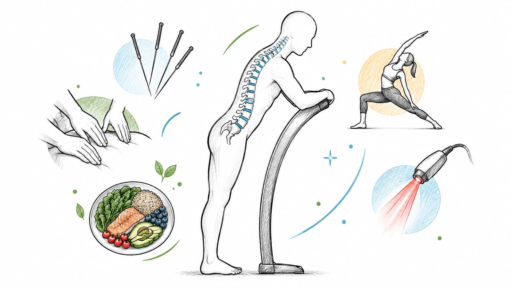
Once pain is less reactive, many clinics add MLS Laser Therapy, SoftWave Therapy, or Acupuncture to calm irritation and make movement easier. Then the emphasis shifts toward Mobility Therapy, Rehabilitation Exercise, and Personal Exercise Plans that teach the spine to tolerate load again.
At Aspen Falls Wellness, one option for this phase-based approach is Spinal Decompression Therapy, often paired with Chiropractic Adjustment, laser-based care, Acupuncture, and home exercise when the exam supports that sequence.
Here's the honest part. Conservative care is first-line, but evidence for every add-on isn't equally strong. A primary-care review reports that about 90% of lumbar disc herniation patients with acute sciatica begin improving within 6 weeks and resolve by 12 weeks with conservative care, while a clinical review reports symptomatic relief in over 90% within 12 weeks in uncomplicated cases. The same evidence summary also notes mixed support for specific add-ons, and an older systematic review found that traction, exercise therapy, and drug therapy were not unequivocally effective for sciatica, while epidural steroids may help selected subgroups, as discussed in this review of nonoperative management.
That doesn't mean every modality is useless. It means clinicians should avoid pretending that every machine or technique fixes every disc. If you're exploring conservative options beyond the standard list, this roundup of uncommon back treatments to try, is a reasonable starting point for understanding the range of approaches patients ask about.
Recovery is rarely linear. Symptoms often calm, flare, then calm again. That's why a timeline matters less as a promise and more as a monitoring framework.
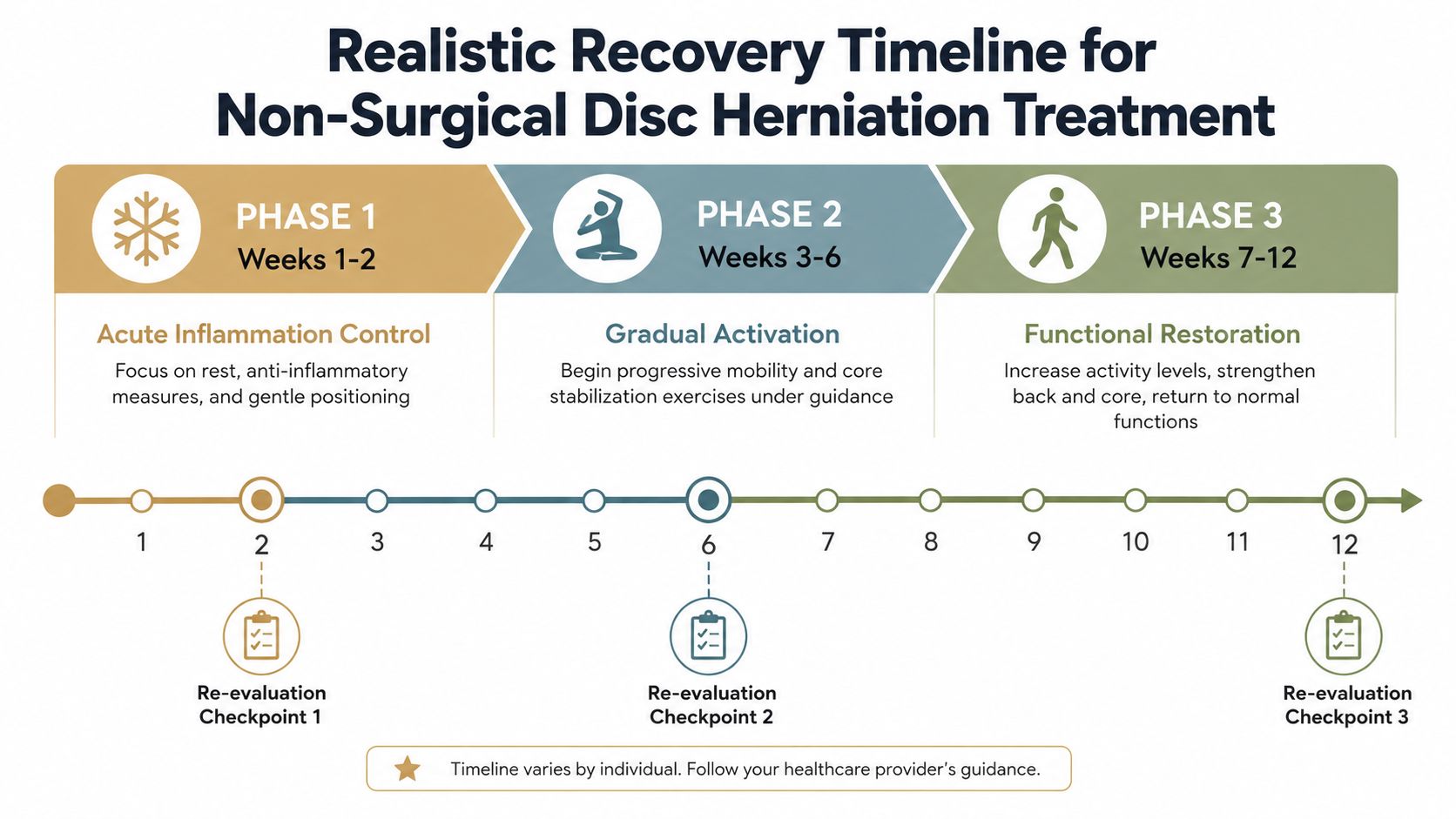
The acute goal is symptom control without deconditioning. Patients usually do best with position changes, short walks, activity modification, and carefully dosed treatment aimed at lowering irritability. This is also the phase where too much bed rest causes trouble.
By this stage, leg pain should start to centralize or at least become less volatile. Motion improves. Sitting tolerance often inches up before people notice dramatic strength changes.
A practical checkpoint at week 2 and another around week 6 should include:
A guideline-based primary care review recommends a 6-week trial of nonoperative management after emergent causes are excluded, and notes that about 90% of patients with acute sciatica from lumbar disc herniation start improving within 6 weeks and resolve by 12 weeks with conservative care, according to the AAFP review on acute lumbar disk pain.
This phase is about building capacity. Heavier strengthening, gait retraining, return-to-work planning, and relapse prevention belong here, not in the first painful week.
For a broader discussion of what patients usually feel at each stage, this herniated disc recovery timeline helps set realistic expectations.
What I watch most closely: pain that retreats from the foot and calf back toward the buttock or low back is often a better sign than a pain number alone.
If the patient is still highly limited at the end of the trial, the question changes from “Can this improve?” to “Is ongoing compression now outlasting safe conservative waiting?”
Most lumbar disc cases are appropriate for conservative care. Some aren't. The danger is assuming every herniation should just be stretched, decompressed, or watched.
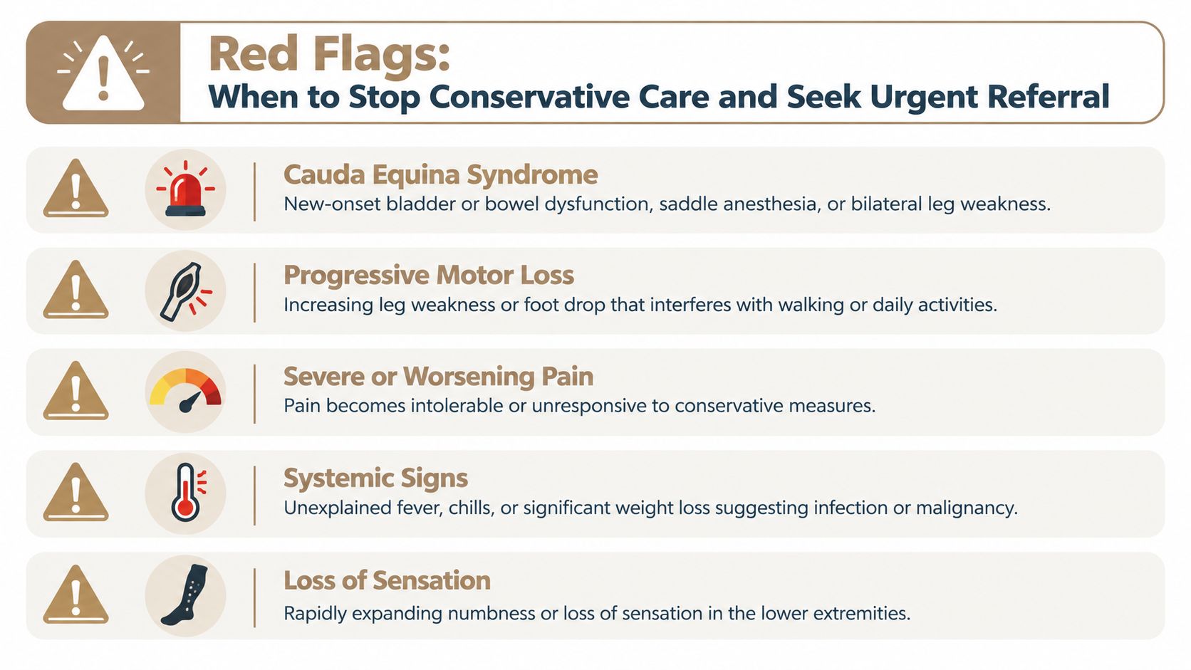
Current guideline synthesis emphasizes that conservative care is first-line mainly when serious neurologic deficits are absent, and also points out that evidence strength varies among noninvasive treatments. In that same broader review, CBT and NSAIDs scored higher than traction and bracing, while TENS wasn't recommended overall, which reinforces the bigger issue: a patient with worsening motor loss needs re-evaluation, not another round of passive care. That decision threshold is outlined well in this Neurospine guideline synthesis.
New weakness changes the conversation. Pain can be monitored. Progressive motor loss demands action.
Some patients don't have cauda equina signs but still shouldn't continue the same plan indefinitely. A patient whose pain remains severe after a structured period of care, or whose strength slips between visits, needs MRI review and likely specialty referral. Another common mistake is treating numbness as harmless because pain is a little better. Sensory change plus declining strength is not reassuring.
Clinic visits matter, but discs don't recover in appointment-sized blocks. The home program fills the gap between treatments and often determines whether gains hold.
In the first stretch, the goal is to calm the nerve without shutting the body down.
Once the pain pattern is clearer, repeated motions can become more specific. If extension centralizes symptoms, extension-based work often becomes the anchor. If extension peripheralizes symptoms, the plan changes. Generic stretching is where many home plans go wrong.
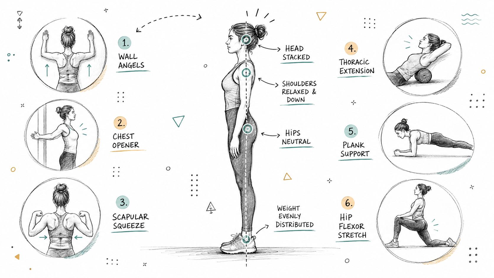
A useful home sequence often includes:
Later, the focus shifts to trunk control and load tolerance. Dead bugs, bird-dogs, side support variations, and controlled hip work usually matter more than high-rep ab circuits. Mobility Therapy and Rehabilitation Exercise should make daily movement easier, not leave the nerve more irritated.
For people who do well with supported core work, these sample Swiss ball back workouts can give ideas for later-stage progression, but only after basic control has returned and the movements fit the patient's presentation.
Home exercise should reinforce in-clinic care. If the program repeatedly drives pain farther down the leg, it needs to be changed, not pushed through.
Nutrition and Nutrition Counseling can also support recovery when inflammation, sleep, and activity tolerance are all part of the picture, though they're adjuncts, not a replacement for mechanical and neurologic management.
The best conservative care doesn't feel scattered. It follows a sequence, measures change, and knows when to stop guessing.
| Integrative Clinic vs Piecemeal Care Key Differences | Integrative Clinic | Piecemeal Care |
|---|---|---|
| Baseline exam | Neurologic and mechanical findings are documented up front | Findings may be split across providers |
| Treatment sequence | Manual care, decompression, pain-modulating therapies, and exercise are staged | Therapies are often added randomly |
| Re-evaluation | Progress is checked at set intervals | Follow-up may depend only on pain complaints |
| Escalation plan | Imaging and referral thresholds are clear | Patients may linger too long in passive care |
| Communication | Providers can align goals inside one plan | The patient often coordinates everything alone |
A well-run integrative setting should be able to answer plain questions. What neurologic deficits are present today? What will be rechecked next visit? When will imaging be ordered? At what point will a spine referral happen? If those answers are vague, the plan is vague.
A major clinical summary notes that most symptomatic lumbar disc herniations resolve within 6 to 8 weeks and are usually managed initially without surgery. It also states that if symptoms persist beyond 6 weeks, epidural steroid injections may be considered for short-term pain relief of about 2 to 4 weeks in selected patients with radiculopathy, according to the NCBI Bookshelf overview of lumbar disc herniation.
That helps define the next step. If leg pain remains severe, weakness progresses, or function barely moves despite consistent care, escalation is appropriate. The goal isn't to avoid surgery at all costs. The goal is to avoid unnecessary surgery while not missing the moment when further delay stops being smart.
Disc herniation treatment without surgery works best when it's active, monitored, and honest about its limits. Real Pain Relief comes from matching the right therapy to the right phase, then changing course when the nerve isn't recovering.
Aspen Falls Wellness provides disc-focused conservative care that can include exam and diagnosis, Chiropractic Care, Spinal Decompression with the DRX 9000, MLS Laser Therapy, SoftWave Therapy, Massage Therapy, Acupuncture, Mobility Therapy, Rehabilitation Exercise, Personal Exercise Plans, and referral when a case needs imaging or surgical input. If you're dealing with sciatica, recurrent disc pain, or a recovery that feels stalled, visit Aspen Falls Wellness to schedule an integrative evaluation and get a plan with clear re-check points and escalation criteria.

Stay updated with the latest chiropractic news and exclusive offers.