Understand lumbar disc herniation and sciatica, including symptoms, diagnosis, conservative treatment options, recovery timelines, and when surgery may be
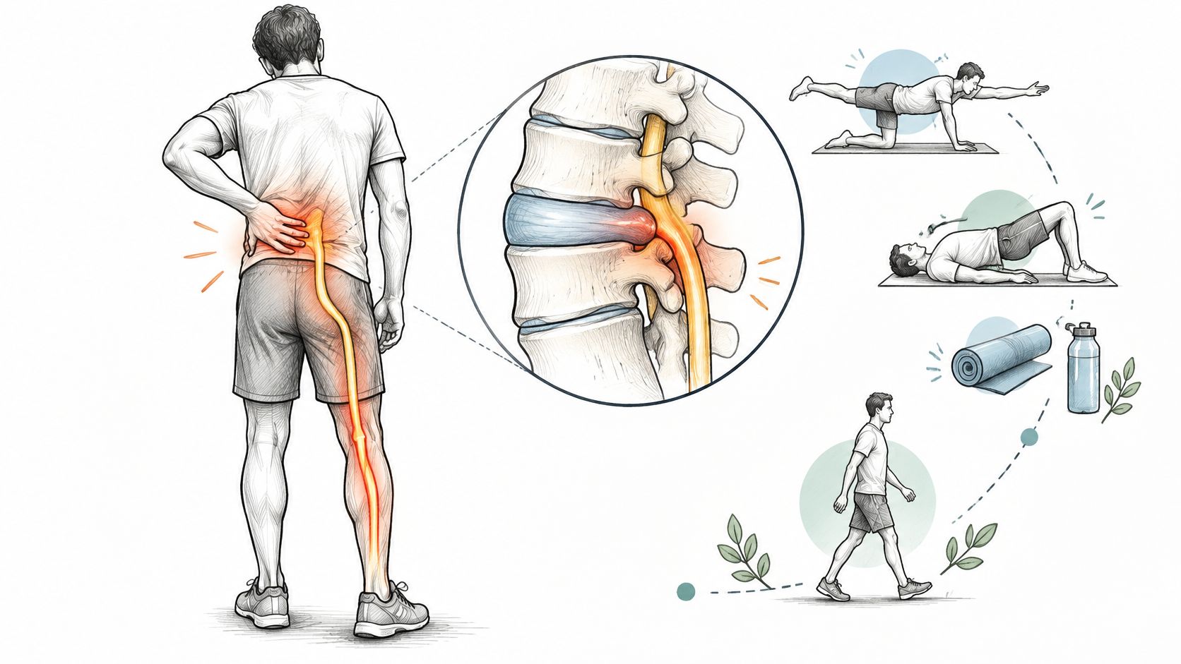
You shift in your chair for the third time in ten minutes, trying to find a position that doesn't send a sharp line of pain from your lower back into your buttock and down your leg. Standing helps for a while. Sitting through a meeting doesn't. You're wondering if this is something you can recover from on your own, or if you're headed for surgery.
That pattern, back discomfort with radiating leg pain that travels below the knee, is the classic experience of lumbar disc herniation and sciatica. You're not imagining how disruptive it is. Sitting at a desk, driving, bending to tie your shoes, and sleeping through the night can all become difficult when a nerve root in your lower back is irritated.
Many people describe the leg symptoms more vividly than the back pain itself. You might feel a burning or electric sensation down the back of your thigh and calf, tingling in your foot, or numbness in a specific patch of skin. Coughing, sneezing, or bending forward can sharpen it. Walking short distances sometimes eases it, while prolonged sitting often makes it worse.
You're also not alone in this. Symptomatic lumbar disc herniation most often affects adults in their third through fifth decades, and men are affected approximately twice as often as women, according to data summarized in a 2025 systematic review of incidence across 87 studies. That same synthesis shows why you'll see very different numbers online. Annual incidence in general population studies ranged from 0.2 to 2.5 cases per 1,000 people when cases were defined by surgery or hospitalization, while one estimate using clinical signs and symptoms reached 13.4 cases per 1,000 people annually.
The gap makes sense once you see it. Far more people develop nerve root symptoms that a clinician can recognize in an office than ever need a hospital stay or an operation. A single prevalence number can't capture that difference.
Your pain being common doesn't make it trivial. It means clinicians have a well tested way to assess it, track it, and decide when to escalate care.
The most useful mindset early on isn't panic and it isn't ignoring the problem either. It's understanding what's irritating the nerve, learning which signs matter most, and following a monitored plan that can change if your trajectory changes. For many, that path starts with conservative care, clear reassessment points, and honest information about what imaging can and can't tell you.
Your spine is a stack of bones called vertebrae, and between each pair sits an intervertebral disc. A helpful analogy is a jelly doughnut. Each disc has a tough outer ring, the annulus fibrosus, and a softer gel like center, the nucleus pulposus. The ring holds the center in place while the disc cushions movement as you bend, twist, and lift.
A herniation happens when that outer ring weakens or tears and some of the inner material pushes out. Think of jelly squeezing through a split in the dough. That displaced material can take up space near a nerve root as it exits the spine, and the chemical irritation around a fresh tear can inflame the nerve even when direct pressure is modest.
Nerve roots from your lower back join to form the sciatic nerve, which carries sensation and muscle signals through your buttock, thigh, calf, and foot. When a root is irritated at the spine, your brain interprets the signal along the whole path of that nerve. That's why the source is in your back but the pain, tingling, or weakness shows up in your leg.
The specific pattern matters. Different roots supply different strips of skin, called dermatomes, and different muscles. Tingling on the top of your foot points to a different root than numbness along the sole. Your clinician uses that map to work backward from your symptoms to the likely level in your spine.
Not all herniations are the same shape. A contained protrusion means the inner material bulges but stays within the outer ring. An extruded herniation means material has pushed through the ring. Counterintuitively, extruded material is often more likely to shrink over time, because your immune system can reach and break it down more easily once it's outside the disc.
This is also why scan wording can mislead you. A large bulge that doesn't touch a nerve may cause no symptoms, while a smaller herniation sitting directly on a root can be very painful. A 2025 review of treatment guidelines for herniated discs with radiculopathy reinforces that first line care is generally nonoperative unless neurological deficits or cauda equina syndrome are present. What matters most isn't the size on the report. It's whether the finding matches your symptoms and examination.
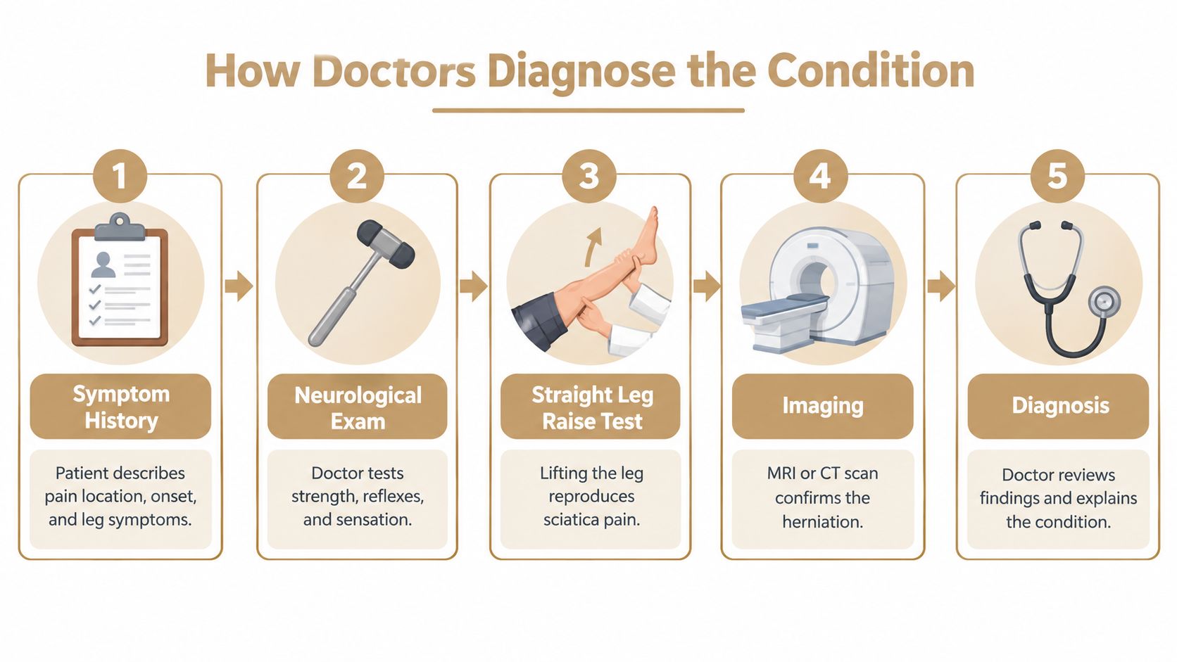
A good diagnosis starts with your story, not a scanner. Your clinician will ask where the pain travels, when it started, what eases or worsens it, and whether you've noticed numbness, weakness, or changes in bowel or bladder control. Those answers narrow the likely nerve root before anyone touches you.
Next comes a focused neurological examination. Your clinician tests muscle strength, reflexes, and sensation in each leg, and may ask you to walk on your heels and toes. The straight leg raise test is also common. You lie on your back while your clinician slowly lifts your leg, and reproducing your familiar leg pain during that stretch supports nerve root irritation.
Many patients expect an MRI at the first visit, so it's confusing when a clinician holds off. The reason is that disc bulges and degenerative changes are common in people with no symptoms at all. Scanning too early can highlight findings that aren't causing your pain, which creates anxiety and sometimes pushes people toward procedures they don't need.
Guidance summarized in a clinical review of lumbar disc herniation imaging decisions recommends avoiding routine CT or MRI at the initial presentation for uncomplicated cases. Imaging becomes appropriate when symptoms persist for approximately 4 to 6 weeks and an epidural injection or surgery is being considered, or sooner if there's severe or progressive neurological deficit or concern for infection, tumor, fracture, or cauda equina syndrome.
NICE guidance on imaging for low back pain with or without sciatica takes the same position. It advises against routinely offering imaging in non specialist settings and says imaging in specialist care should happen only when the result is likely to change management. If you're weighing scan types, this overview of MRI compared with X ray for back pain explains what each test shows well.
Practical rule: A scan earns its place when the result would change what you and your clinician do next.
When you do get imaging, look for one word in the interpretation, concordant. That means the herniation sits at the level and side that match your leg symptoms and examination findings. A concordant MRI carries far more decision value than an incidental bulge found on the opposite side from your pain.
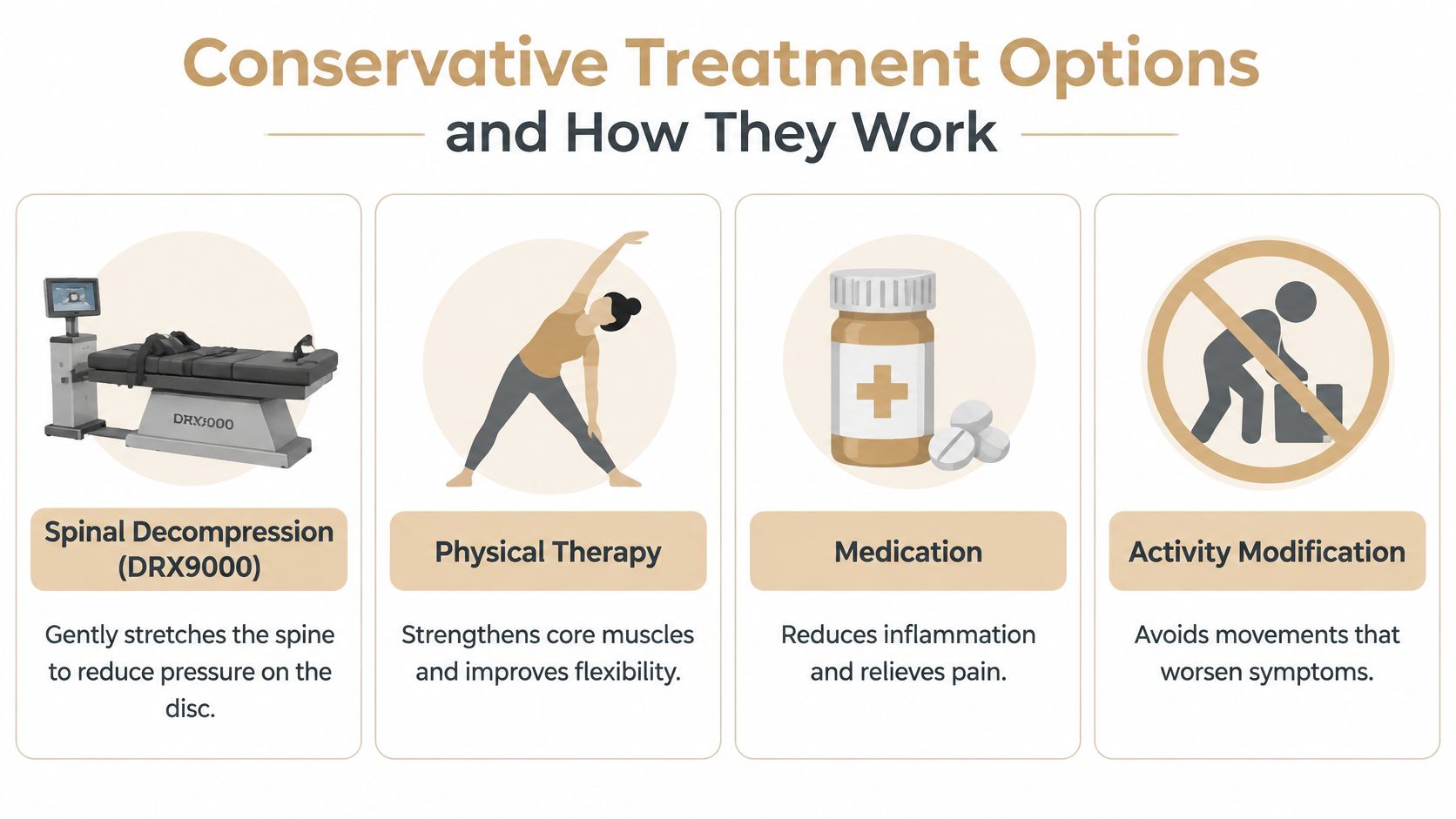
Conservative care isn't one treatment. It's a combination of approaches, each targeting a different part of the problem, and the mix should reflect how irritable your symptoms are and what functions you're trying to recover.
Spinal decompression with the DRX 9000 uses a motorized table to apply gentle, controlled traction to your spine in cycles. The goal is to reduce pressure within the disc and create more space around the irritated nerve root. You lie secured on the table while the device stretches and relaxes in a measured rhythm, and the sensation is generally tolerable rather than painful.
Honesty matters here. A systematic review of motorized nonsurgical spinal decompression studies analyzed 10 studies, including seven randomized controlled trials, and found that six of the seven reported no difference between motorized decompression and comparison treatment. Three uncontrolled studies reported pain reductions of 77% to 86%, but without control groups those results are vulnerable to placebo effects and natural recovery. Decompression is best framed as one option within a monitored plan, judged by your pain, function, and neurological status, not as a guaranteed disc correction.
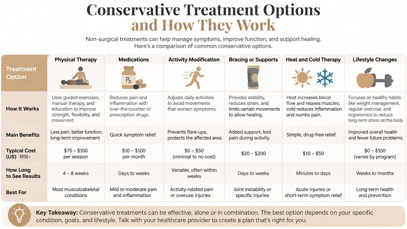
Passive treatments can calm symptoms, but personal exercise plans and rehabilitation exercise are what rebuild capacity. The Agency for Healthcare Research and Quality summary on noninvasive back pain treatments reports that exercise may be effective for radicular low back pain, although the strength of evidence is low. Your program might combine biomechanical, aerobic, or mind body approaches based on your needs and preferences, progressed as your leg symptoms allow.
Practices such as Aspen Falls Wellness deliver this kind of combined model, coordinating chiropractic care, decompression, laser therapy, acupuncture, mobility therapy, and home exercise under one team. Whichever setting you choose, ask how progress will be reevaluated and what happens if you aren't improving.
The question underneath most others is simple. How long will this take? The natural history of this condition is more reassuring than typical patients expect, and it shapes why clinicians usually start conservatively.
Clinical reviews summarized in an overview of lumbar disc herniation natural history and resorption report that roughly 85% to 90% of people with an acute herniated lumbar disc experience symptom relief within 6 to 12 weeks without surgery. That doesn't mean every week feels better than the last. Recovery is often uneven, with a good stretch followed by a flare after a long drive or an awkward lift, even while the overall trend improves.

Disc material that has pushed out can be gradually broken down and reabsorbed, a process called spontaneous resorption. The same source above cites a meta analysis reporting resorption in 66.6% of cases, and a 2024 systematic review of 2,199 patients found resorption in 1,684 cases, or 76.6%, occurring mainly during the first 3 to 6 months of conservative treatment. Extruded herniations resorb more readily than contained protrusions, which is one reason a dramatic looking early scan doesn't predict a dramatic outcome.
Pain often settles before the scan would change. Evidence reviewed in NICE guidance on assessment and noninvasive treatment for low back pain and sciatica notes that pain decreases within 3 months in approximately 87% of patients treated without surgery. For a fuller walkthrough of phases and milestones, see this guide to how long herniated disc recovery typically takes.
Judge your plan by trajectory over weeks, not by whether today hurts more than yesterday.
It's natural to treat surgery and conservative care as opposite, permanent choices. The evidence paints a more time dependent picture, and understanding that trade off helps you decide without fear driving the conversation.
A 2025 evidence based guideline comparing surgery with nonsurgical care for lumbar disc herniation found that surgery produced substantially greater leg pain reduction at 6 months, with a mean between group difference of -2.40 points. By 2 years, the difference was -0.95 points and no longer statistically significant. In plain terms, surgery tends to relieve leg pain faster for appropriately selected patients, but long term outcomes converge for many people.
That pattern reframes the question. Surgery isn't mainly about whether you can eventually improve without it. It's about how much disability you're living with now, how long you can reasonably tolerate it, and what risks you're willing to accept to recover sooner.
Discectomy, the operation that removes the herniated fragment pressing on the root, is the procedure most often discussed for this condition. It's aimed primarily at leg pain from nerve compression, not at general back ache or at making a scan look normal.
A severe looking MRI by itself doesn't make you a surgical candidate, and mild imaging doesn't disqualify you if your function is deteriorating. Bring four things to that conversation. Your symptom duration, your functional limits at work and home, your neurological examination findings, and your personal priorities around speed of relief versus surgical risk. A good clinician will also separate routine radiating pain, which can be monitored, from progressive weakness or bladder changes, which can't wait. If those urgent signs appear, the decision stops being elective.
Most cases of lumbar disc herniation and sciatica can be managed conservatively and monitored over weeks. A small set of findings changes that completely. Learning to separate routine nerve pain from these warning signs protects your long term nerve function.
Other situations also warrant urgent evaluation, including back pain with fever that could suggest infection, pain after significant trauma that could mean fracture, or a history and presentation raising concern for tumor. These aren't sciatica specific, but they change what your clinician needs to rule out first. This summary of low back pain red flags is worth saving on your phone so you can check symptoms quickly if something changes.
Call urgently for new weakness, saddle numbness, or bladder changes. Don't wait for your next scheduled appointment to mention them.
Acting quickly matters because nerves compressed severely, especially in cauda equina syndrome, can sustain lasting damage if decompression is delayed. If you develop these symptoms, contact your clinician immediately or go to an emergency department and describe the neurological changes specifically, not just the pain. Stable tingling in one dermatome, by contrast, is common in ordinary sciatica and usually doesn't signal an emergency, though you should still report any change at your next visit.
Recovery works best as an active, monitored process rather than passive waiting. You don't need to do everything at once. You need a clear starting point, a way to measure whether it's working, and agreed moments to change course.
Start with an evaluation that includes a neurological examination, not just a conversation about pain. Ask your clinician three direct questions. Which nerve root do my symptoms point to? What findings would make you order imaging or refer me? And how will we measure progress at each visit? Clear answers signal a structured plan.
Your core program will usually combine education, activity guidance, exercise, and one or more passive modalities matched to your irritability. Stay as active as your leg symptoms allow, favor frequent short walks over bed rest, and modify sitting with regular position changes. Add nutrition counseling or mobility therapy where they support your broader goals, and keep any single treatment accountable to functional change.
Keep a brief weekly log you can bring to appointments. Rate your leg pain, record your sitting and walking tolerance in minutes, note heel and toe walking ability, and write down sleep quality and one daily task that's getting easier or harder. Review the log every two weeks rather than daily, because real trends are easier to see at that distance.
Set reassessment points in advance. If you're clearly improving by six weeks, continuing conservative care makes sense. If you're plateaued with disabling leg pain and concordant imaging, that's the moment for a surgical opinion, and seeking one doesn't commit you to an operation. If red flag symptoms appear at any point, skip the schedule and seek urgent care.
You deserve a team that explains findings in plain language, coordinates your chiropractic adjustment, soft tissue work, decompression, laser therapy, acupuncture, and exercise rather than stacking them randomly, and refers you onward when your needs fall outside its scope. Advocating for yourself means asking what each treatment is for, when you'll reevaluate it, and what success should look like in your daily life, not just on a scan.
If radiating leg pain is limiting your work, sleep, or daily movement, Aspen Falls Wellness offers coordinated non surgical care in Salt Lake City and Sandy, including chiropractic adjustment, DRX 9000 spinal decompression, MLS Laser Therapy, SoftWave Therapy, acupuncture, massage, and personalized exercise plans built around a thorough neurological evaluation. Schedule an assessment to learn which nerve root is involved, whether imaging would change your plan, and how your progress will be tracked week by week.

Stay updated with the latest chiropractic news and exclusive offers.