Explore an evidence-based lumbar disc herniation treatment protocol. Learn about DRX9000 decompression, phased conservative care, and referral criteria.
A sharp pain in the low back can turn into burning pain down the buttock and leg within minutes. You may be unable to sit, sleep, or put on your shoes, and an MRI report may make the situation sound even more alarming. The important first question isn't whether the disc looks dramatic. It's whether your symptoms indicate an emergency, a routine nerve irritation that can settle, or a problem that needs closer monitoring and escalation.
A sound lumbar disc herniation treatment protocol follows that order. It starts with neurological triage, uses active conservative care when appropriate, tracks function rather than relying on imaging alone, and defines clear thresholds for specialist referral. The aim isn't to delay necessary treatment. It's to avoid unnecessary intervention while acting quickly when nerve function is at risk.
The first appointment should establish what the pain is doing to the nerve, not just where the MRI shows a disc abnormality. A clinician will review the onset of symptoms, leg pain distribution, numbness, weakness, aggravating movements, and any changes in bowel or bladder function. The examination should include walking, strength, sensation, reflexes, spinal movement, and relevant nerve tension tests.
Most acute lumbar disc herniations with radicular symptoms don't require immediate surgery. A recent systematic review of clinical guidelines reports that more than 85% of patients with acute herniated lumbar discs and radiculopathy recover over time, while StatPearls notes that many symptomatic cases improve within 6 to 8 weeks. Conservative treatment is often recommended for 6 weeks to 2 months before surgery is considered when there are no emergency findings, as summarized in the systematic review of lumbar disc herniation guidelines.
That recovery pattern reflects more than pain tolerance. Disc material can gradually regress, inflammation can settle, and the irritated nerve can regain normal function as the mechanical and chemical irritation decreases. A large finding on an MRI therefore doesn't automatically determine the treatment outcome.
A practical initial pathway usually looks like this:
Clinical rule: The MRI describes anatomy. The neurological examination and the patient's changing function determine urgency.
For patients without red flags, the conservative-first approach isn't a promise that every case will resolve on the same schedule. Some people improve quickly, while others have persistent leg pain, restricted movement, or weakness that requires a more active plan. The practical value of conservative care is that it gives the body an opportunity to recover while clinicians monitor whether the nerve is improving, stable, or deteriorating.
Patients who want a detailed discussion of nonsurgical options can review this guide to disc herniation treatment without surgery. The next step should still be an individualized examination, especially when numbness or weakness is present.
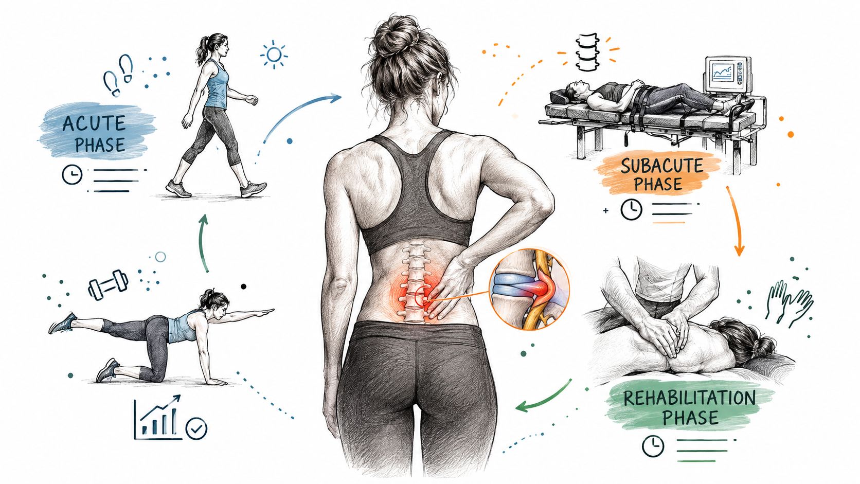
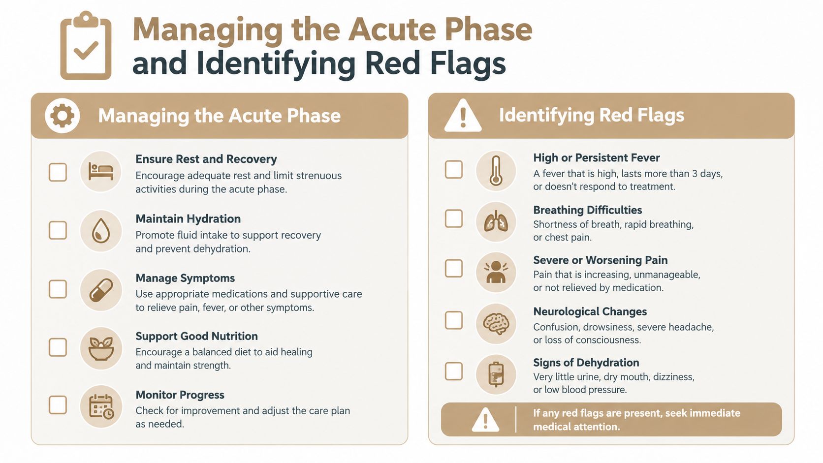
The acute phase is about controlling aggravating forces without shutting the body down. Major patient guidance recommends limiting activity for about 2 to 3 days, walking as tolerated, and avoiding routine prolonged bed rest or traction, as outlined by the American Association of Neurological Surgeons guidance on herniated discs. That doesn't mean pushing through severe leg pain. It means changing the movements that provoke symptoms while preserving manageable activity.
Sitting for long periods, repeated forward bending, heavy lifting, and twisting may aggravate an inflamed nerve. Short walks, frequent position changes, and supported rest often provide a better balance than staying in bed. Analgesics or NSAIDs may be appropriate for some patients, but medication choice depends on medical history, kidney and stomach health, other prescriptions, and guidance from a qualified medical professional.
A conservative plan may include:
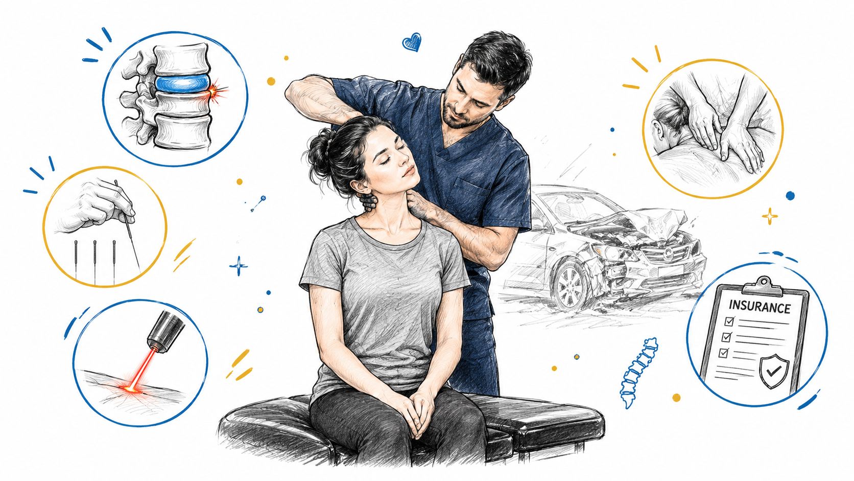
An initial chiropractic evaluation shouldn't be treated as a substitute for neurological triage. An adjustment may be considered when it fits the examination and the patient's tolerance, but forceful treatment of an unstable or worsening neurological presentation isn't appropriate. The same principle applies to acupuncture, muscle stimulation, and other pain-relief modalities. They can support comfort and movement, but they don't replace urgent medical assessment when nerve function is declining.
Seek urgent evaluation if you develop new or progressive motor weakness, such as a foot that repeatedly slaps the floor, an inability to lift the foot, or a marked loss of strength when pushing off. Bowel or bladder dysfunction, including new loss of control or difficulty emptying, is an emergency concern. Saddle anesthesia, meaning numbness around the groin, genitals, buttocks, or inner thighs, also requires immediate assessment.
A clinician should also respond promptly to rapidly worsening neurological symptoms, severe unrelenting pain with concerning examination findings, or other signs suggesting a serious condition rather than uncomplicated disc irritation. In these situations, the correct next step may be urgent imaging and referral to a spine or emergency specialist, not another round of routine conservative treatment.
When severe pain begins to settle but radicular symptoms continue, the plan should become more targeted. The patient may still need activity modification, but the clinical emphasis shifts toward restoring movement, reducing protective muscle guarding, and testing whether the nerve tolerates progressive loading.
Standard physical therapy and advanced device-based care can serve different purposes. Exercise-based rehabilitation builds control, strength, and confidence through active movement. Manual therapy may improve restricted motion or reduce muscular guarding. A device such as the DRX9000 is used as a non-surgical spinal decompression approach, with treatment selected according to the examination, symptom behavior, and tolerance rather than the MRI image alone.
The proposed rationale for motorized decompression is controlled unloading of the affected spinal segment. Clinicians use the treatment to reduce compressive loading and create conditions that may make movement more comfortable. It should be integrated into a broader plan, not presented as a replacement for neurological monitoring, rehabilitation, or surgical referral when red flags appear.
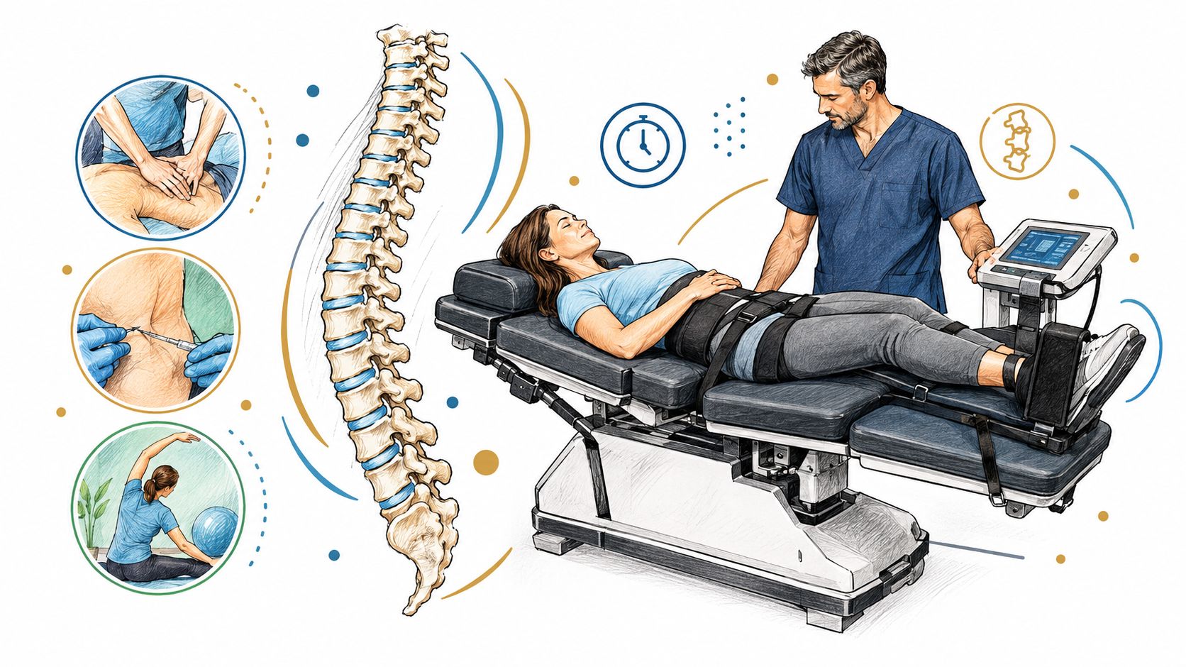
Exercise-based rehabilitation is the foundation for rebuilding tolerance. A practitioner may begin with positions that reduce symptoms, gentle neural mobility, pelvic control, and trunk endurance, then progress toward lifting and work-specific movements. The correct exercise isn't necessarily the one that feels hardest. It's the one that improves function without causing a lasting increase in leg symptoms.
Spinal decompression with the DRX9000 may be considered when persistent disc-related pain limits participation in active care. A Johns Hopkins-associated cohort used a 6-week DRX9000 treatment protocol and reported disc height increasing from 7.5 mm to 8.8 mm, while reported low back pain decreased from 6.2 to 1.6. Both changes were reported as statistically significant at p<0.001 in the published study of nonsurgical spinal decompression. Those findings are encouraging, but a cohort result doesn't establish that every patient will experience the same response.
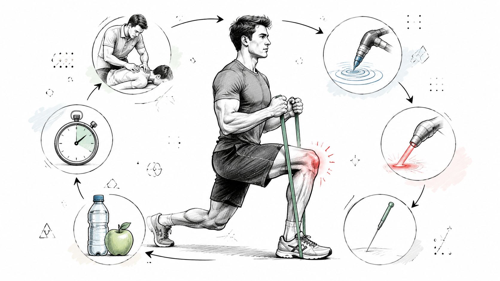
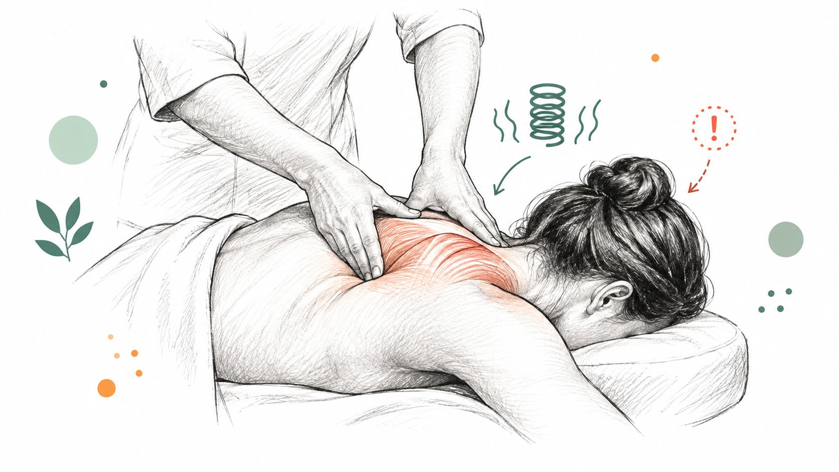
MLS Laser Therapy can be used as a local, non-invasive modality when inflammation and tissue sensitivity make movement difficult. It may be paired with massage therapy to address guarding in the lumbar, gluteal, and hip tissues. The purpose is practical, to improve comfort enough for the patient to move, exercise, and sleep, rather than to make a device the entire treatment plan.
The clinical discussion of spinal decompression therapy should always include candid expectations. Decompression may be a reasonable component for selected patients, but it can't correct progressive weakness or cauda equina syndrome. A clinician should reassess pain distribution, strength, sensation, and activity tolerance throughout the program.
Recovery becomes more durable when treatment changes from symptom control to capacity building. A patient who can walk farther but still lacks hip mobility, trunk control, or confidence with bending isn't finished. The rehabilitation phase should connect hands-on care with exercises the patient can perform independently.
Chiropractic adjustment may be used to address joint restriction and support realignment when the examination identifies a mechanical limitation and the technique is well tolerated. The adjustment should serve a functional goal, such as improving comfortable rotation or reducing protective stiffness, rather than being repeated without measuring change.
SoftWave Therapy can be incorporated as a tissue-stimulation modality during rehabilitation, while MLS Laser Therapy may be used when local sensitivity or inflammation continues to interfere with movement. Massage Therapy can reduce compensatory muscle tension in the back, hips, and legs. These services work best when each has a defined role and the team can remove, change, or progress a modality based on the patient's response.
A coordinated program may combine:
The key is sequencing. A patient with irritable sciatica may first need pain relief and comfortable positions. Once symptoms stabilize, the same patient may benefit more from active strengthening than from continued passive treatment. Mobility therapy and posture retraining then help transfer those gains into sitting, lifting, walking, and work activities.
A multidisciplinary model also improves communication. The chiropractor can identify a strength change, the mobility specialist can modify an exercise, the massage therapist can note a new pattern of guarding, and the acupuncturist can adjust pain-modulation care. That information should flow into one plan with regular reassessment, not remain in separate treatment silos.
A broader multidisciplinary pain management approach is especially useful when pain has affected sleep, work, exercise, and mood at the same time. The goal is not to keep adding treatments indefinitely. It's to use the right intervention for the current barrier, then progress toward independence.
A large herniation on an MRI doesn't automatically mean you need an operation. Imaging findings must match the pain pattern, neurological examination, and functional limitations. Spontaneous disc resorption is common enough that current guidance generally supports an initial conservative trial when there is no cauda equina syndrome, severe progressive deficit, or disabling radicular pain that fails to improve.
The more useful question is, “What is changing?” Leg pain that is becoming less intense, traveling a shorter distance, or appearing less often suggests a different pathway from leg pain that remains constant and disabling. Back pain may persist while nerve pain improves, so clinicians should track both rather than treating the MRI as the only outcome.
At each review, document:
For persistent radicular pain without deterioration, a guideline review reports that surgery is usually considered after 6 to 12 weeks of conservative treatment. The same review states that cauda equina syndrome requires emergency decompression within 24 to 48 hours, while moderate or severe paresis, graded MRC 3/5 or less, should prompt surgery within 3 days. These thresholds are summarized in the guideline review on timing for lumbar disc herniation surgery.
A separate review of guidelines emphasizes that early surgery can be preferable for certain motor deficits, while nonoperative care remains appropriate for many patients without neurological red flags. That distinction matters. “Wait 6 weeks” isn't a safe instruction for someone whose strength is rapidly declining, and “operate because the MRI is large” isn't evidence-based for someone whose function is steadily improving.
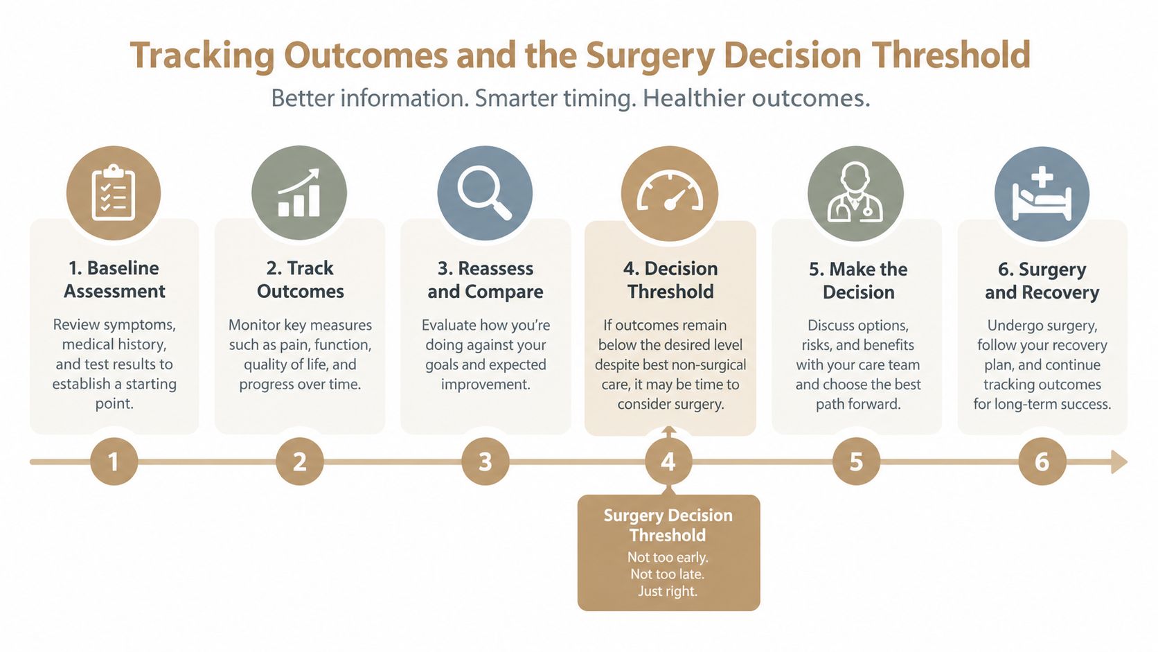
Referral to a spine surgeon is appropriate when progressive weakness appears, emergency symptoms develop, or disabling leg pain persists despite a well-delivered conservative program. It can also be appropriate when the patient understands the trade-off and wants a specialist opinion because faster relief has become a priority.
Randomized evidence makes that trade-off clear. In a BMJ trial, 141 patients were assigned to early surgery and 142 to prolonged conservative care. 125 patients, or 89%, in the early-surgery group underwent microdiscectomy, while 62 patients, or 44%, in the conservative group eventually required surgery. Early surgery produced faster leg-pain relief, but disability outcomes were similar by one year and remained comparable over two years, as reported in the BMJ trial of early surgery and prolonged conservative care.
That doesn't make surgery good or bad in isolation. It makes timing a shared clinical decision based on neurological safety, symptom severity, recovery direction, and the patient's goals.
Returning to normal activity shouldn't mean waiting until every sensation disappears. It means restoring enough strength, mobility, coordination, and confidence to meet the demands of the activity safely. A patient who can walk comfortably but can't hinge, lift, or rotate under control needs more preparation before returning to heavier work or sport.
Start with the take-home rehabilitation plan. Perform the prescribed exercises consistently, keep the range controlled, and monitor what happens later that day and the following morning. A mild muscular response can be acceptable, but increasing leg pain, spreading numbness, or new weakness calls for reassessment rather than a harder workout.
Active adults should consider a Golf Movement Screening before returning to a rotational sport. The assessment can identify restrictions in hip rotation, thoracic mobility, balance, or swing mechanics that may cause the lumbar spine to absorb more stress than necessary. Similar movement-specific screening can be adapted for lifting, running, cycling, or other physical demands.
Use practical ergonomics at home and work. Bring loads close to the body, avoid twisting while lifting, divide awkward loads when possible, and alternate prolonged sitting with standing or walking. If a flare occurs, return briefly to the last comfortable level of activity instead of stopping all movement.
A durable recovery plan also accounts for future injuries. Aspen Falls Wellness provides evaluation, chiropractic care, spinal decompression with the DRX 9000, SoftWave Therapy, MLS Laser Therapy, massage, acupuncture, mobility care, nutrition counseling, and rehabilitation exercise for patients who need a coordinated nonsurgical pathway.
If persistent sciatica, numbness, or activity-limiting low back pain is disrupting your routine, schedule an evaluation with Aspen Falls Wellness. The team can assess neurological status, build a phased lumbar disc herniation treatment protocol, and refer you for specialist care when your symptoms fall outside conservative management.

Stay updated with the latest chiropractic news and exclusive offers.