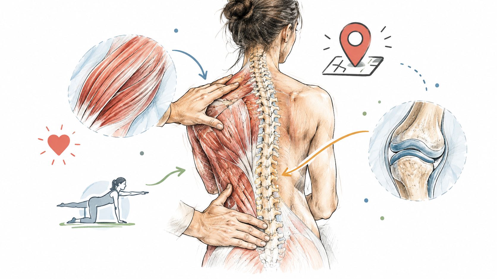
Learn about spinal stenosis and degenerative disc disease, including symptoms, causes, and treatment options to manage your condition effectively.
A person in their late sixties may notice the change in a grocery store first. A mile-long walk that once felt routine becomes a slow trip down one aisle. Halfway through, they stop, brace against a shelf, and wait for the heaviness and tingling in their legs to settle. Sitting would help, but they'd rather keep moving, one careful step at a time.
That pattern often leads to an MRI and two intimidating phrases, spinal stenosis and degenerative disc disease. The diagnosis should come with more than a list of abnormal findings. You deserve to understand what's happening inside your spine, why standing can hurt more than sitting, and what realistic options exist before anyone mentions surgery. If you're also looking for practical habits that may support back comfort, this guide to relieve lower back pain naturally offers additional general lifestyle context.
The two questions underneath almost every appointment are simple: What's causing these symptoms, and do I have to go under the knife to fix them? In many cases, the answer requires matching your symptoms and function to your imaging, then giving structured conservative care a fair trial.
The grocery-store example is more than a story about aging. It captures a recognizable pattern called neurogenic claudication, in which standing or walking brings on leg heaviness, aching, cramping, tingling, or weakness. Sitting or leaning forward often provides relief because that position changes the shape of the lower spine and gives crowded nerve tissue more room.
A person may describe it as needing to stop at every bench, shortening their walking route, or choosing a shopping cart because leaning forward makes movement easier. The limitation isn't always severe back pain. Sometimes the legs feel tired, unreliable, or strangely disconnected from the effort required to walk.
Lumbar spinal stenosis is common at a population level. A systematic review estimated the pooled prevalence of clinically diagnosed lumbar spinal stenosis at about 11% in the general population, while radiologic narrowing appeared in people without symptoms as well, at about 11% among asymptomatic people and about 38% in the general population, depending on the setting and definition used. The figures come from a review available through the published systematic review of lumbar spinal stenosis prevalence.
That matters because an MRI can describe a crowded space without proving that the space is responsible for every symptom. Some people with notable narrowing continue to walk, work, and sleep reasonably well. Others have less dramatic-looking findings but substantial limitations because the nerves are sensitive, the surrounding joints are irritated, or multiple problems are occurring together.
Non-surgical care isn't a promise to restore a younger spine or erase every structural change. It's a way to reduce irritation, improve movement, build capacity, and monitor whether your function is changing in the right direction.
You'll learn how disc degeneration and stenosis connect, what symptoms deserve attention, how clinicians interpret imaging, and when a conservative plan should give way to specialist or surgical evaluation.
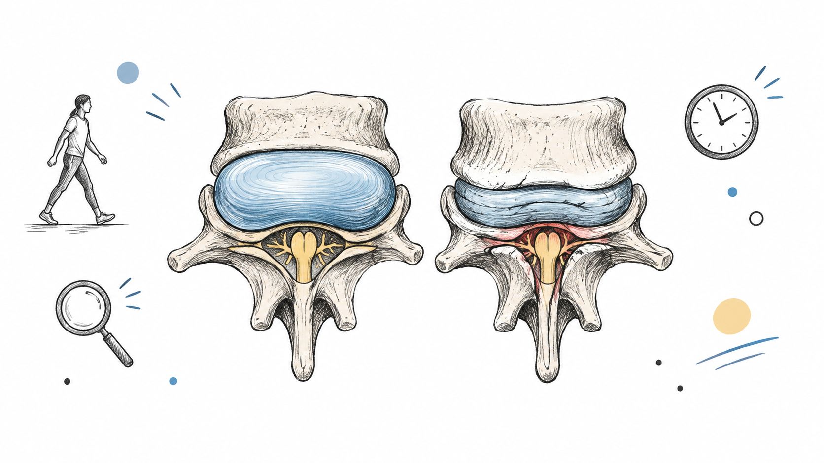
Start with two simple images. Think of a spinal disc as a shock absorber between two bones, and think of the nerve pathway as a doorway that must remain open enough for traffic to pass. Degenerative disc disease affects the shock absorber. Spinal stenosis describes the narrowing around the doorway.
A healthy disc has a hydrated inner portion and a strong outer ring. Over time, the disc can lose water, develop fibrotic changes, and become shorter. MRI may show reduced signal and disc-space collapse. The disc then distributes load less effectively, so the facet joints and other posterior parts of the spinal segment may carry more stress. The mechanisms are described in this review of degenerative disc disease and disc biomechanics.
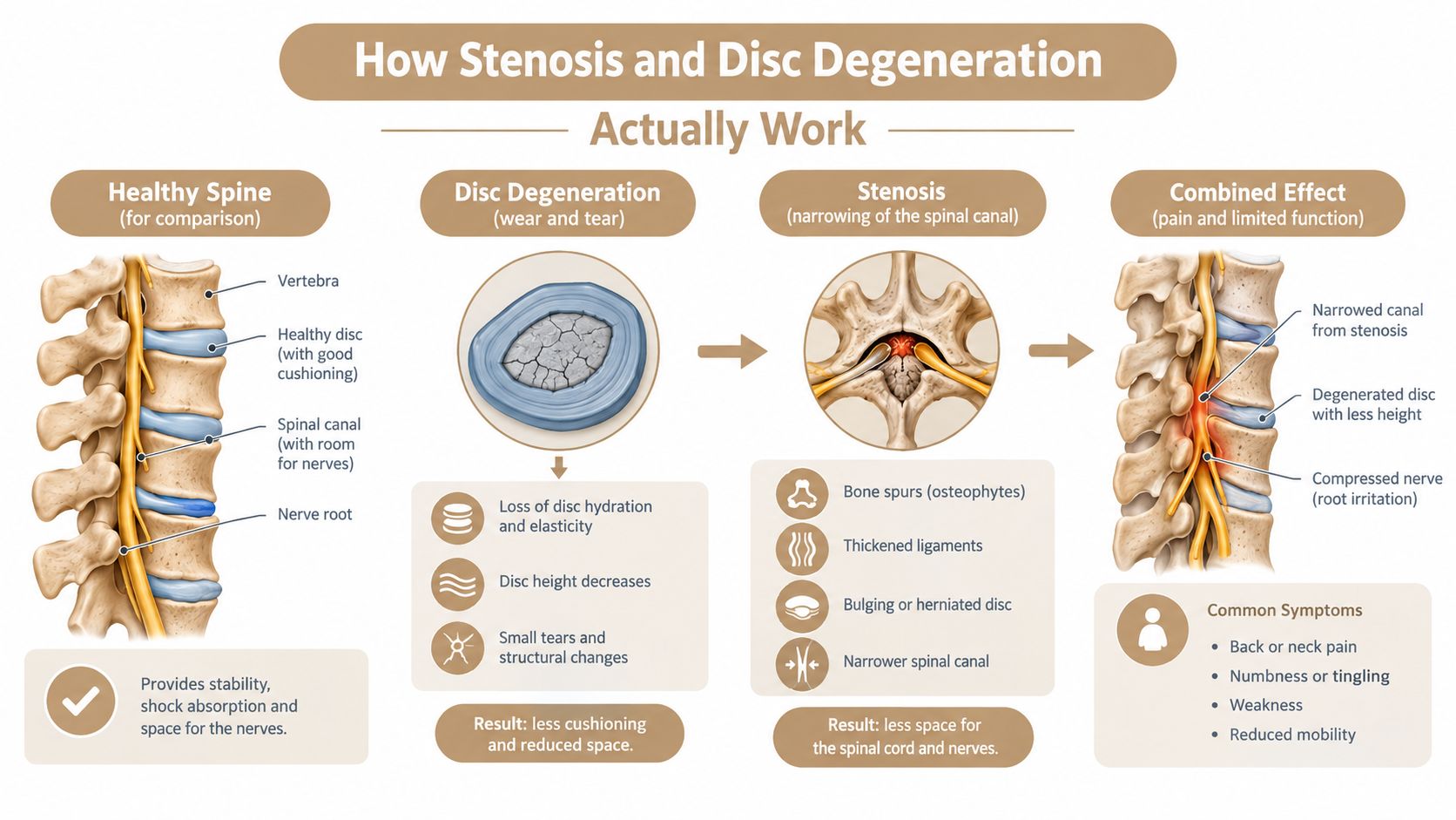
As a disc loses height, the nearby joints and ligaments experience a changed mechanical environment. A disc bulge, facet-joint enlargement, thickened ligament, or bone overgrowth can occupy more of the canal, lateral recess, or neural foramen. Those are the spaces where the spinal cord or nerve roots travel.
That's why the diagnoses often overlap. DDD describes the condition of the disc, while stenosis describes the reduced space available for neural structures. One person can have disc degeneration without symptomatic nerve compression. Another can have stenosis driven by several age-related changes, with the disc playing only one part.
The term “degenerative disc disease” can also mislead people. It doesn't mean an infection or an automatically progressive catastrophe. It usually refers to age-linked structural change with possible mechanical consequences. Imaging findings become clinically important when they fit the person's symptoms, examination, and functional limitations.
A major review of pain-free people found disc degeneration on imaging in 37% of asymptomatic 20-year-olds, rising steadily to 96% of asymptomatic 80-year-olds. The age-related pattern is summarized in this systematic review of imaging findings in asymptomatic people. A disc can look worn without being the sole cause of pain.
A clinician doesn't diagnose symptomatic stenosis by measuring one line on an MRI report. They ask what brings on your symptoms, where those symptoms travel, what positions change them, and how your walking and balance have changed.
Neurogenic claudication usually has a posture-dependent pattern. Standing upright, extending the lower back, or walking downhill may increase leg symptoms. Sitting, bending slightly forward, or leaning on a cart may reduce them. Flexion can create more room in a narrowed pathway, while extension can make that pathway tighter.
Radicular pain follows an irritated nerve root and may travel from the back or buttock into the thigh, calf, or foot. Numbness and tingling can follow a similar distribution, although people often describe these sensations less precisely. Weakness deserves careful examination, especially when a leg repeatedly gives way, a person catches a toe, or stair climbing becomes noticeably harder.
Vascular claudication can also cause leg discomfort with walking, so posture alone doesn't settle the diagnosis. A clinician may consider circulation, pulses, medical history, reflexes, sensation, muscle strength, gait, and how quickly symptoms ease after stopping. The point is to identify the source rather than assume every walking limitation comes from the spine.
MRI remains important. A best-practice guideline describes stenosis as narrowing of the central canal, lateral recess, or neural foramen, and identifies MRI as the preferred imaging method for diagnosis. It also recommends initial conservative care unless fixed or progressive neurological deficits are present, as outlined in this guideline on lumbar spinal stenosis diagnosis and management.
For additional education about how different imaging tests answer different questions, review this comparison of MRI and X-ray for back pain.
| MRI Finding | What It Often Means | How It Correlates With Symptoms |
|---|---|---|
| Disc dehydration or reduced MRI signal | The disc has lost water content and normal structure | It may contribute to mechanical pain, but it can also appear without symptoms |
| Reduced disc height | The disc space has narrowed | It can reduce foraminal room, especially when nerve symptoms match the same level |
| Central canal narrowing | Less space exists around the nerve structures | The relationship with pain and disability is often weak unless the clinical pattern fits |
| Foraminal narrowing | The opening for a nerve root is smaller | Radiating pain, numbness, or weakness may be meaningful when they follow that nerve's pathway |
| Severe central stenosis | Marked narrowing is present | The most severe narrowing may have stronger symptom associations, but treatment still depends on examination and function |
One meta-analysis found central canal stenosis on imaging in 14.0% of asymptomatic people versus 59.5% of symptomatic people, while disc degeneration appeared in 34.4% of asymptomatic people versus 57.4% of symptomatic people. Those findings are reported in this meta-analysis of imaging abnormalities in symptomatic and asymptomatic people. The practical lesson is straightforward: an MRI should inform treatment, not dictate it by itself.
Not every ache requires an emergency visit, and not every tolerable symptom should be ignored. A useful appointment starts with an accurate description of what you can still do, what has changed, and whether the trend is stable or worsening.
Use the table as a conversation tool, not as a self-diagnosis. A clinician still needs to assess strength, sensation, reflexes, gait, and any relevant medical causes.
| Category | What It Looks Like | What To Do |
|---|---|---|
| Tolerable symptoms | Occasional stiffness, mild aching that improves after changing position, or brief numbness that resolves within minutes | Arrange a routine evaluation and track triggers, walking tolerance, and recovery time |
| Functional limits | A shrinking walking distance, difficulty standing for 10 to 15 minutes, or regular reliance on a cart, railing, or nearby seat | Request a structured examination, discuss imaging when indicated, and begin care that measures functional progress |
| Warning signs | Progressive leg weakness, foot drop, saddle anesthesia, bowel or bladder changes, unremitting night pain, or rapid escalation | Seek urgent medical evaluation. New or worsening neurological changes may require prompt imaging or specialist referral |
The distinction between pain and function is important. Pain may fluctuate with sleep, activity, stress, and position, while declining walking tolerance can reveal a more meaningful change in nerve or movement capacity.
Practical rule: A stable symptom that responds to position is different from a neurological deficit that is steadily taking away function.
Don't wait for a routine chiropractic or primary-care appointment if you develop new loss of bowel or bladder control, numbness in the saddle area, rapidly worsening gait, or clear progressive weakness. Those signs need prompt medical attention.
Non-surgical care shouldn't be framed as doing nothing until surgery becomes unavoidable. It's an active process that combines symptom control, movement retraining, strength, education, and regular reassessment.
A long-term natural-history study of degenerative lumbar spinal stenosis found that most patients with neurogenic claudication remained ambulatory and didn't develop motor deficits or sphincter dysfunction. The authors concluded that conservative management is reasonable for people with tolerable symptoms and low functional expectations, particularly when certain anatomical risk factors are absent, as described in this long-term study of degenerative lumbar spinal stenosis. That doesn't make every case safe to observe. It does challenge the idea that stenosis leads to surgery.
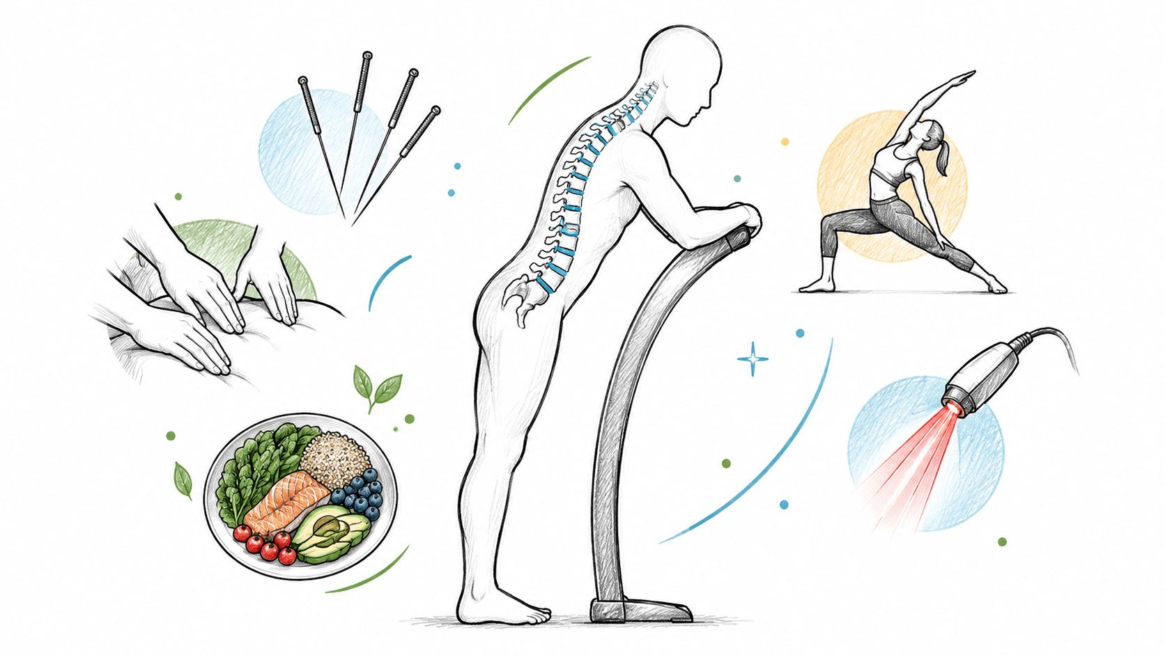
At Aspen Falls Wellness, an integrated plan may include several services, selected according to the examination and the person's tolerance:
The combination makes clinical sense because each modality addresses a different barrier. Reducing pain may make movement possible, while movement and strength help preserve the gains after a passive treatment ends.
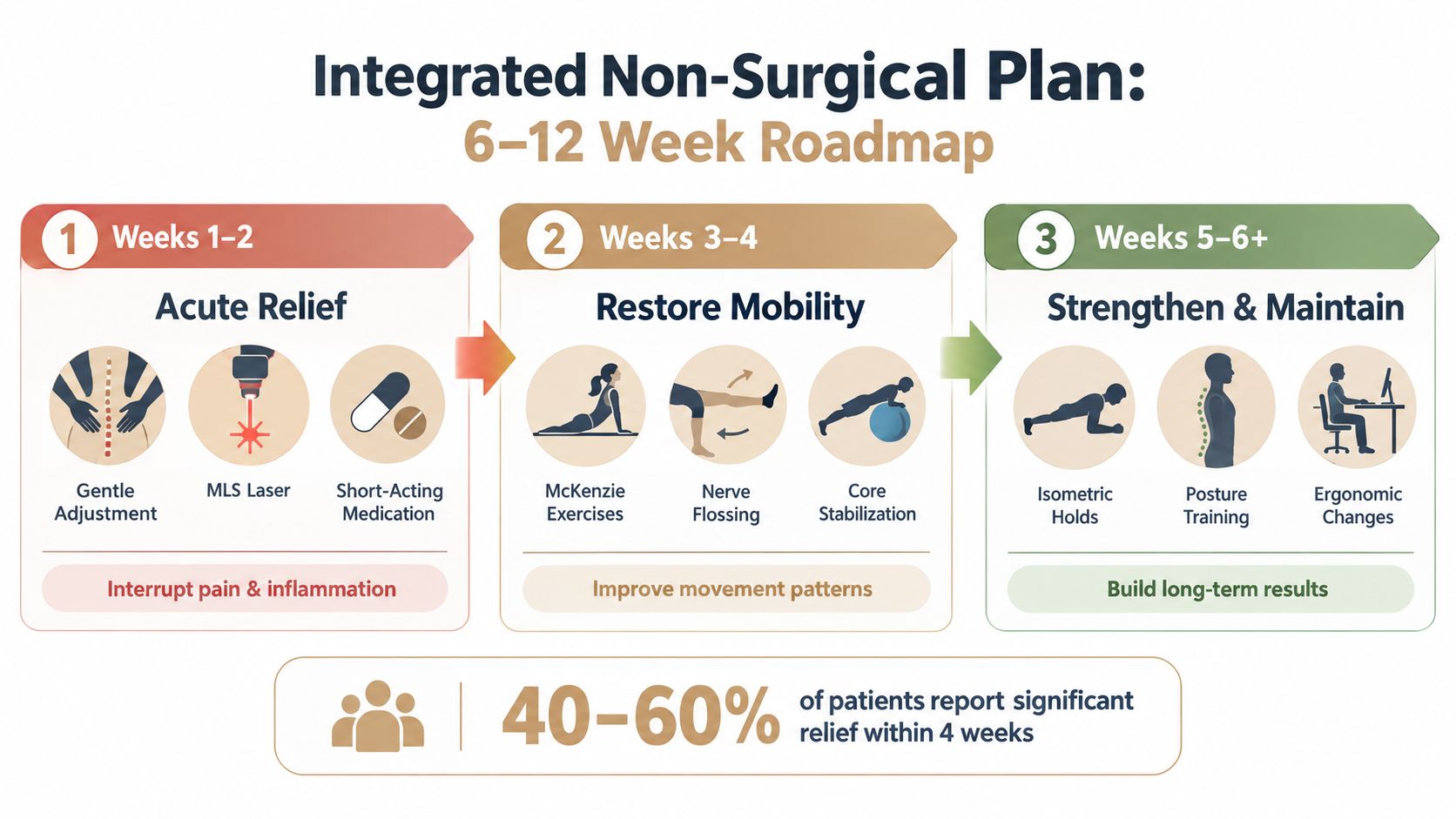
A realistic plan should feel specific enough that you know what the next visit is meant to accomplish. The schedule must remain individualized, but a 6 to 12 week framework can provide useful checkpoints rather than leaving care open-ended.
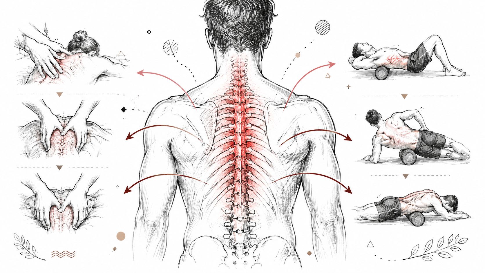
During the first 1 to 2 weeks, the priority is often reducing irritability. Gentle Chiropractic Adjustment, MLS Laser Therapy, Massage Therapy, acupuncture, and comfortable positioning may be used according to the examination. The clinician also establishes a baseline, such as walking distance, standing tolerance, sleep disruption, leg symptoms, and strength.
Medication decisions belong with the prescribing clinician. A short medication course may sometimes create a window for movement, but medication isn't the foundation of the rehabilitation plan.
The next phase may introduce Spinal Decompression with the DRX 9000 for appropriate disc-related presentations, alongside mobility therapy and early stabilization exercises. The plan shouldn't chase a sensation of being “realigned” if walking, standing, and daily tasks aren't improving.
People often ask what a decompression appointment feels like. The answer varies by protocol and tolerance, but treatment should be comfortable enough to avoid provoking a sustained flare. You can learn more about the general approach through this spinal decompression therapy overview.
Later care emphasizes Rehabilitation Exercise, gait training, posture retraining, and endurance. Nutrition and Nutrition Counseling may support broader health goals, while Massage Therapy, Acupuncture, SoftWave Therapy, or MLS Laser Therapy can remain available when pain or soft-tissue guarding interferes with activity.
At the week 4 and week 8 checkpoints, the important questions are measurable: Can you stand longer? Walk farther? Recover faster? Sleep more consistently? If progress is flat, the plan should change. If neurological signs are worsening, referral takes priority over continuing the same treatment.
Three assumptions commonly push people toward poor decisions.
A report that uses words such as “severe” can feel like a direct explanation for every symptom. Yet reviews of lumbar stenosis imaging report little to no consistent correlation between the measured degree of narrowing and pain, disability, or function. One study found a strong association only with the most severe central narrowing, with an odds ratio of 15.5 and a 95% confidence interval from 1.4 to 164.9, as summarized in this review of imaging and symptom correlation in lumbar spinal stenosis.
The better question is whether the finding matches your posture-dependent leg symptoms, neurological examination, and walking limitation.
The natural-history evidence doesn't support an automatic march from diagnosis to operation. Many people with tolerable symptoms remain ambulatory and can pursue conservative care, particularly when they don't have progressive neurological loss.
That doesn't mean surgery is wrong. It means the decision should reflect function, neurological status, symptom trajectory, anatomy, and response to a structured trial.
Rest can calm an acute flare, but prolonged inactivity often leaves the legs and trunk less prepared for ordinary demands. A person may feel safer sitting, then discover that standing tolerance has fallen further when they try to resume normal activity.
The target isn't perfect imaging. It's safer movement, better function, and a clear response to care.
Conservative care has reached its limit when the plan is well structured, progress has been tracked, and function remains stalled or neurological status worsens. Pain reduction matters, but it isn't the only outcome. Walking distance, standing tolerance, gait quality, strength, sensation, sleep, and daily independence should all inform the decision.
A legitimate non-surgical trial often spans 6 to 12 weeks, with individualized treatment and documented reassessment. That may include Chiropractic Care, Chiropractic Adjustment, Spinal Decompression, Mobility Therapy, Massage Therapy, Acupuncture, Muscle Stimulation, SoftWave Therapy, MLS Laser Therapy, and Rehabilitation Exercise when clinically appropriate. It may also include Personal Exercise Plans, nutrition support, and activity changes that help you maintain progress outside the clinic.
If improvement has stalled without these red flags, request a re-evaluation rather than either giving up too early or continuing indefinitely without a measurable goal. Ask whether updated imaging, a second clinical opinion, or a surgical consultation would clarify the next step. A consultation doesn't obligate you to have surgery. It gives you better information.
For a focused discussion of non-surgical options, see this guide to stenosis treatment without surgery.
Aspen Falls Wellness offers individualized Chiropractic Care, DRX 9000 Spinal Decompression, MLS Laser Therapy, SoftWave Therapy, Massage Therapy, Acupuncture, and Rehabilitation Exercise for people managing stenosis, degenerative disc disease, and related pain. Visit Aspen Falls Wellness to request an evaluation and discuss a plan built around your symptoms, neurological findings, and functional goals.

Stay updated with the latest chiropractic news and exclusive offers.